Magnesium »
PDB 2p8b-2ppb »
2pa4 »
Magnesium in PDB 2pa4: Crystal Structure of Udp-Glucose Pyrophosphorylase From Corynebacteria Glutamicum in Complex with Magnesium and Udp-Glucose
Enzymatic activity of Crystal Structure of Udp-Glucose Pyrophosphorylase From Corynebacteria Glutamicum in Complex with Magnesium and Udp-Glucose
All present enzymatic activity of Crystal Structure of Udp-Glucose Pyrophosphorylase From Corynebacteria Glutamicum in Complex with Magnesium and Udp-Glucose:
2.7.7.9;
2.7.7.9;
Protein crystallography data
The structure of Crystal Structure of Udp-Glucose Pyrophosphorylase From Corynebacteria Glutamicum in Complex with Magnesium and Udp-Glucose, PDB code: 2pa4
was solved by
H.M.Holden,
J.B.Thoden,
with X-Ray Crystallography technique. A brief refinement statistics is given in the table below:
| Resolution Low / High (Å) | 20.00 / 2.00 |
| Space group | C 1 2 1 |
| Cell size a, b, c (Å), α, β, γ (°) | 173.400, 47.600, 161.500, 90.00, 102.70, 90.00 |
| R / Rfree (%) | n/a / n/a |
Magnesium Binding Sites:
The binding sites of Magnesium atom in the Crystal Structure of Udp-Glucose Pyrophosphorylase From Corynebacteria Glutamicum in Complex with Magnesium and Udp-Glucose
(pdb code 2pa4). This binding sites where shown within
5.0 Angstroms radius around Magnesium atom.
In total 8 binding sites of Magnesium where determined in the Crystal Structure of Udp-Glucose Pyrophosphorylase From Corynebacteria Glutamicum in Complex with Magnesium and Udp-Glucose, PDB code: 2pa4:
Jump to Magnesium binding site number: 1; 2; 3; 4; 5; 6; 7; 8;
In total 8 binding sites of Magnesium where determined in the Crystal Structure of Udp-Glucose Pyrophosphorylase From Corynebacteria Glutamicum in Complex with Magnesium and Udp-Glucose, PDB code: 2pa4:
Jump to Magnesium binding site number: 1; 2; 3; 4; 5; 6; 7; 8;
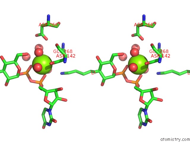
Magnesium binding site 1 out of 8 in 2pa4
Go back to
Magnesium binding site 1 out
of 8 in the Crystal Structure of Udp-Glucose Pyrophosphorylase From Corynebacteria Glutamicum in Complex with Magnesium and Udp-Glucose

Mono view

Stereo pair view
Mono view
Stereo pair view
A full contact list of Magnesium with other atoms in the Mg binding
site number 1 of Crystal Structure of Udp-Glucose Pyrophosphorylase From Corynebacteria Glutamicum in Complex with Magnesium and Udp-Glucose within 5.0Å range:
|
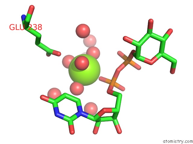
Magnesium binding site 2 out of 8 in 2pa4
Go back to
Magnesium binding site 2 out
of 8 in the Crystal Structure of Udp-Glucose Pyrophosphorylase From Corynebacteria Glutamicum in Complex with Magnesium and Udp-Glucose

Mono view
Stereo pair view
Mono view
Stereo pair view
A full contact list of Magnesium with other atoms in the Mg binding
site number 2 of Crystal Structure of Udp-Glucose Pyrophosphorylase From Corynebacteria Glutamicum in Complex with Magnesium and Udp-Glucose within 5.0Å range:
|
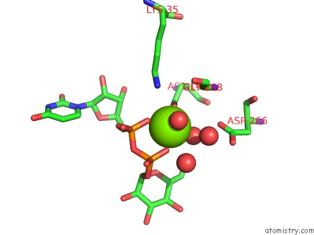
Magnesium binding site 3 out of 8 in 2pa4
Go back to
Magnesium binding site 3 out
of 8 in the Crystal Structure of Udp-Glucose Pyrophosphorylase From Corynebacteria Glutamicum in Complex with Magnesium and Udp-Glucose

Mono view

Stereo pair view
Mono view
Stereo pair view
A full contact list of Magnesium with other atoms in the Mg binding
site number 3 of Crystal Structure of Udp-Glucose Pyrophosphorylase From Corynebacteria Glutamicum in Complex with Magnesium and Udp-Glucose within 5.0Å range:
|
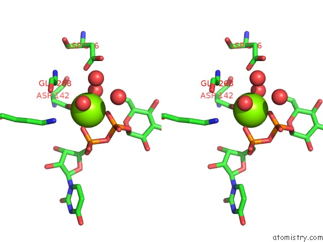
Magnesium binding site 4 out of 8 in 2pa4
Go back to
Magnesium binding site 4 out
of 8 in the Crystal Structure of Udp-Glucose Pyrophosphorylase From Corynebacteria Glutamicum in Complex with Magnesium and Udp-Glucose

Mono view

Stereo pair view
Mono view
Stereo pair view
A full contact list of Magnesium with other atoms in the Mg binding
site number 4 of Crystal Structure of Udp-Glucose Pyrophosphorylase From Corynebacteria Glutamicum in Complex with Magnesium and Udp-Glucose within 5.0Å range:
|
Magnesium binding site 5 out of 8 in 2pa4
Go back to
Magnesium binding site 5 out
of 8 in the Crystal Structure of Udp-Glucose Pyrophosphorylase From Corynebacteria Glutamicum in Complex with Magnesium and Udp-Glucose

Mono view

Stereo pair view
Mono view
Stereo pair view
A full contact list of Magnesium with other atoms in the Mg binding
site number 5 of Crystal Structure of Udp-Glucose Pyrophosphorylase From Corynebacteria Glutamicum in Complex with Magnesium and Udp-Glucose within 5.0Å range:
|
Magnesium binding site 6 out of 8 in 2pa4
Go back to
Magnesium binding site 6 out
of 8 in the Crystal Structure of Udp-Glucose Pyrophosphorylase From Corynebacteria Glutamicum in Complex with Magnesium and Udp-Glucose

Mono view

Stereo pair view
Mono view
Stereo pair view
A full contact list of Magnesium with other atoms in the Mg binding
site number 6 of Crystal Structure of Udp-Glucose Pyrophosphorylase From Corynebacteria Glutamicum in Complex with Magnesium and Udp-Glucose within 5.0Å range:
|
Magnesium binding site 7 out of 8 in 2pa4
Go back to
Magnesium binding site 7 out
of 8 in the Crystal Structure of Udp-Glucose Pyrophosphorylase From Corynebacteria Glutamicum in Complex with Magnesium and Udp-Glucose
Mono view

Stereo pair view
Mono view
Stereo pair view
A full contact list of Magnesium with other atoms in the Mg binding
site number 7 of Crystal Structure of Udp-Glucose Pyrophosphorylase From Corynebacteria Glutamicum in Complex with Magnesium and Udp-Glucose within 5.0Å range:
|
Magnesium binding site 8 out of 8 in 2pa4
Go back to
Magnesium binding site 8 out
of 8 in the Crystal Structure of Udp-Glucose Pyrophosphorylase From Corynebacteria Glutamicum in Complex with Magnesium and Udp-Glucose
Mono view
Stereo pair view
Mono view
Stereo pair view
A full contact list of Magnesium with other atoms in the Mg binding
site number 8 of Crystal Structure of Udp-Glucose Pyrophosphorylase From Corynebacteria Glutamicum in Complex with Magnesium and Udp-Glucose within 5.0Å range:
|
Reference:
J.B.Thoden,
H.M.Holden.
Active Site Geometry of Glucose-1-Phosphate Uridylyltransferase. Protein Sci. V. 16 1379 2007.
ISSN: ISSN 0961-8368
PubMed: 17567737
DOI: 10.1110/PS.072864707
Page generated: Sun Aug 10 12:53:19 2025
ISSN: ISSN 0961-8368
PubMed: 17567737
DOI: 10.1110/PS.072864707
Last articles
Mg in 6QPHMg in 6QV0
Mg in 6QUZ
Mg in 6QUY
Mg in 6QUX
Mg in 6QUW
Mg in 6QUV
Mg in 6QUU
Mg in 6QUS
Mg in 6QTN