Magnesium »
PDB 2p8b-2ppb »
2plj »
Magnesium in PDB 2plj: Crystal Structure of Lysine/Ornithine Decarboxylase Complexed with Putrescine From Vibrio Vulnificus
Enzymatic activity of Crystal Structure of Lysine/Ornithine Decarboxylase Complexed with Putrescine From Vibrio Vulnificus
All present enzymatic activity of Crystal Structure of Lysine/Ornithine Decarboxylase Complexed with Putrescine From Vibrio Vulnificus:
4.1.1.17; 4.1.1.18;
4.1.1.17; 4.1.1.18;
Protein crystallography data
The structure of Crystal Structure of Lysine/Ornithine Decarboxylase Complexed with Putrescine From Vibrio Vulnificus, PDB code: 2plj
was solved by
J.Lee,
E.J.Goldsmith,
M.A.Phillips,
with X-Ray Crystallography technique. A brief refinement statistics is given in the table below:
| Resolution Low / High (Å) | 19.72 / 1.70 |
| Space group | P 21 21 21 |
| Cell size a, b, c (Å), α, β, γ (°) | 82.074, 88.683, 111.843, 90.00, 90.00, 90.00 |
| R / Rfree (%) | 18.1 / 21.3 |
Magnesium Binding Sites:
The binding sites of Magnesium atom in the Crystal Structure of Lysine/Ornithine Decarboxylase Complexed with Putrescine From Vibrio Vulnificus
(pdb code 2plj). This binding sites where shown within
5.0 Angstroms radius around Magnesium atom.
In total only one binding site of Magnesium was determined in the Crystal Structure of Lysine/Ornithine Decarboxylase Complexed with Putrescine From Vibrio Vulnificus, PDB code: 2plj:
In total only one binding site of Magnesium was determined in the Crystal Structure of Lysine/Ornithine Decarboxylase Complexed with Putrescine From Vibrio Vulnificus, PDB code: 2plj:
Magnesium binding site 1 out of 1 in 2plj
Go back to
Magnesium binding site 1 out
of 1 in the Crystal Structure of Lysine/Ornithine Decarboxylase Complexed with Putrescine From Vibrio Vulnificus
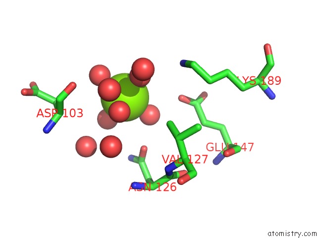
Mono view
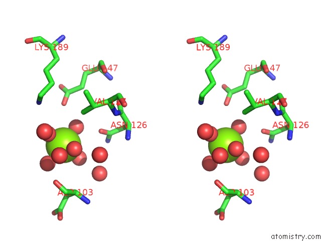
Stereo pair view
Mono view
Stereo pair view
A full contact list of Magnesium with other atoms in the Mg binding
site number 1 of Crystal Structure of Lysine/Ornithine Decarboxylase Complexed with Putrescine From Vibrio Vulnificus within 5.0Å range:
|
Reference:
J.Lee,
A.J.Michael,
D.Martynowski,
E.J.Goldsmith,
M.A.Phillips.
Phylogenetic Diversity and the Structural Basis of Substrate Specificity in the Beta/Alpha-Barrel Fold Basic Amino Acid Decarboxylases. J.Biol.Chem. V. 282 27115 2007.
ISSN: ISSN 0021-9258
PubMed: 17626020
DOI: 10.1074/JBC.M704066200
Page generated: Wed Aug 14 02:09:44 2024
ISSN: ISSN 0021-9258
PubMed: 17626020
DOI: 10.1074/JBC.M704066200
Last articles
Zn in 9J0NZn in 9J0O
Zn in 9J0P
Zn in 9FJX
Zn in 9EKB
Zn in 9C0F
Zn in 9CAH
Zn in 9CH0
Zn in 9CH3
Zn in 9CH1