Magnesium »
PDB 2p8b-2ppb »
2pn3 »
Magnesium in PDB 2pn3: Crystal Structure of Hepatitis C Virus Ires Subdomain Iia
Protein crystallography data
The structure of Crystal Structure of Hepatitis C Virus Ires Subdomain Iia, PDB code: 2pn3
was solved by
Q.Zhao,
Q.Han,
C.R.Kissinger,
T.Hermann,
P.A.Thompson,
with X-Ray Crystallography technique. A brief refinement statistics is given in the table below:
| Resolution Low / High (Å) | 45.00 / 2.90 |
| Space group | P 43 21 2 |
| Cell size a, b, c (Å), α, β, γ (°) | 48.945, 48.945, 120.942, 90.00, 90.00, 90.00 |
| R / Rfree (%) | 22.9 / 28.3 |
Other elements in 2pn3:
The structure of Crystal Structure of Hepatitis C Virus Ires Subdomain Iia also contains other interesting chemical elements:
| Bromine | (Br) | 2 atoms |
Magnesium Binding Sites:
The binding sites of Magnesium atom in the Crystal Structure of Hepatitis C Virus Ires Subdomain Iia
(pdb code 2pn3). This binding sites where shown within
5.0 Angstroms radius around Magnesium atom.
In total 2 binding sites of Magnesium where determined in the Crystal Structure of Hepatitis C Virus Ires Subdomain Iia, PDB code: 2pn3:
Jump to Magnesium binding site number: 1; 2;
In total 2 binding sites of Magnesium where determined in the Crystal Structure of Hepatitis C Virus Ires Subdomain Iia, PDB code: 2pn3:
Jump to Magnesium binding site number: 1; 2;
Magnesium binding site 1 out of 2 in 2pn3
Go back to
Magnesium binding site 1 out
of 2 in the Crystal Structure of Hepatitis C Virus Ires Subdomain Iia
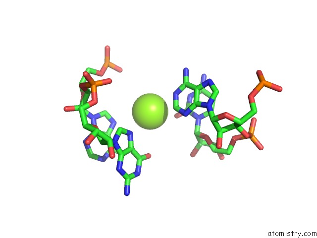
Mono view
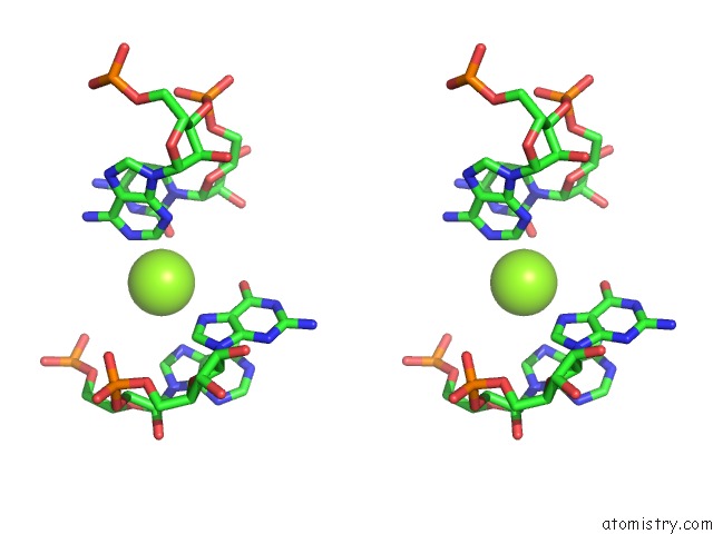
Stereo pair view
Mono view
Stereo pair view
A full contact list of Magnesium with other atoms in the Mg binding
site number 1 of Crystal Structure of Hepatitis C Virus Ires Subdomain Iia within 5.0Å range:
|
Magnesium binding site 2 out of 2 in 2pn3
Go back to
Magnesium binding site 2 out
of 2 in the Crystal Structure of Hepatitis C Virus Ires Subdomain Iia
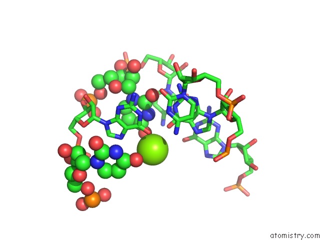
Mono view
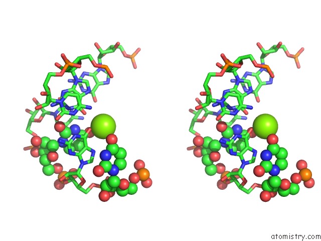
Stereo pair view
Mono view
Stereo pair view
A full contact list of Magnesium with other atoms in the Mg binding
site number 2 of Crystal Structure of Hepatitis C Virus Ires Subdomain Iia within 5.0Å range:
|
Reference:
Q.Zhao,
Q.Han,
C.R.Kissinger,
T.Hermann,
P.A.Thompson.
Structure of Hepatitis C Virus Ires Subdomain Iia. Acta Crystallogr.,Sect.D V. 64 436 2008.
ISSN: ISSN 0907-4449
PubMed: 18391410
DOI: 10.1107/S0907444908002011
Page generated: Wed Aug 14 02:12:34 2024
ISSN: ISSN 0907-4449
PubMed: 18391410
DOI: 10.1107/S0907444908002011
Last articles
Zn in 9J0NZn in 9J0O
Zn in 9J0P
Zn in 9FJX
Zn in 9EKB
Zn in 9C0F
Zn in 9CAH
Zn in 9CH0
Zn in 9CH3
Zn in 9CH1