Magnesium »
PDB 2ppq-2q0e »
2pup »
Magnesium in PDB 2pup: Structures of 5-Methylthioribose Kinase Reveal Substrate Specificity and Unusual Mode of Nucleotide Binding
Enzymatic activity of Structures of 5-Methylthioribose Kinase Reveal Substrate Specificity and Unusual Mode of Nucleotide Binding
All present enzymatic activity of Structures of 5-Methylthioribose Kinase Reveal Substrate Specificity and Unusual Mode of Nucleotide Binding:
2.7.1.100;
2.7.1.100;
Protein crystallography data
The structure of Structures of 5-Methylthioribose Kinase Reveal Substrate Specificity and Unusual Mode of Nucleotide Binding, PDB code: 2pup
was solved by
S.-Y.Ku,
with X-Ray Crystallography technique. A brief refinement statistics is given in the table below:
| Resolution Low / High (Å) | 46.34 / 2.60 |
| Space group | P 21 21 2 |
| Cell size a, b, c (Å), α, β, γ (°) | 213.930, 83.290, 51.420, 90.00, 90.00, 90.00 |
| R / Rfree (%) | 21.1 / 27.4 |
Magnesium Binding Sites:
The binding sites of Magnesium atom in the Structures of 5-Methylthioribose Kinase Reveal Substrate Specificity and Unusual Mode of Nucleotide Binding
(pdb code 2pup). This binding sites where shown within
5.0 Angstroms radius around Magnesium atom.
In total 4 binding sites of Magnesium where determined in the Structures of 5-Methylthioribose Kinase Reveal Substrate Specificity and Unusual Mode of Nucleotide Binding, PDB code: 2pup:
Jump to Magnesium binding site number: 1; 2; 3; 4;
In total 4 binding sites of Magnesium where determined in the Structures of 5-Methylthioribose Kinase Reveal Substrate Specificity and Unusual Mode of Nucleotide Binding, PDB code: 2pup:
Jump to Magnesium binding site number: 1; 2; 3; 4;
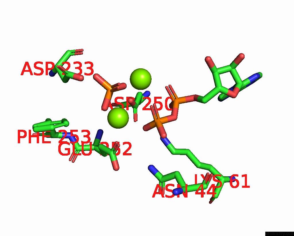
Magnesium binding site 1 out of 4 in 2pup
Go back to
Magnesium binding site 1 out
of 4 in the Structures of 5-Methylthioribose Kinase Reveal Substrate Specificity and Unusual Mode of Nucleotide Binding
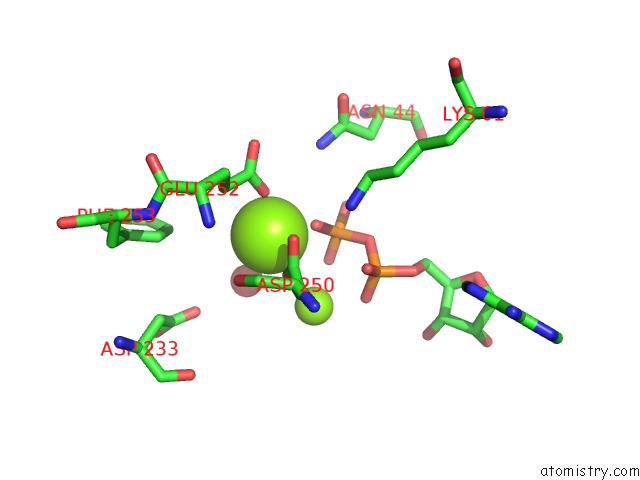
Mono view
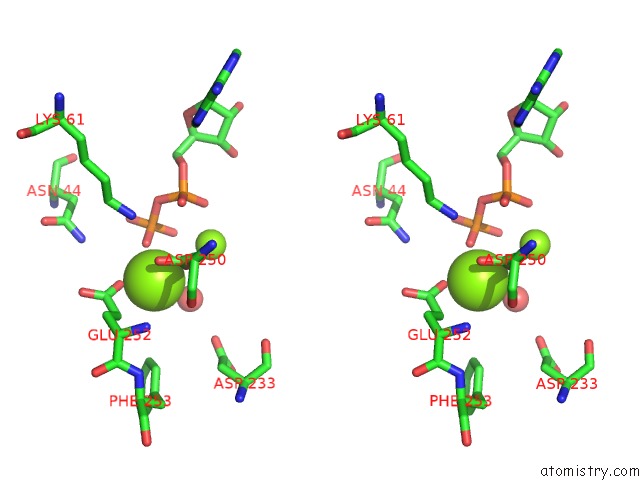
Stereo pair view
Mono view
Stereo pair view
A full contact list of Magnesium with other atoms in the Mg binding
site number 1 of Structures of 5-Methylthioribose Kinase Reveal Substrate Specificity and Unusual Mode of Nucleotide Binding within 5.0Å range:
|
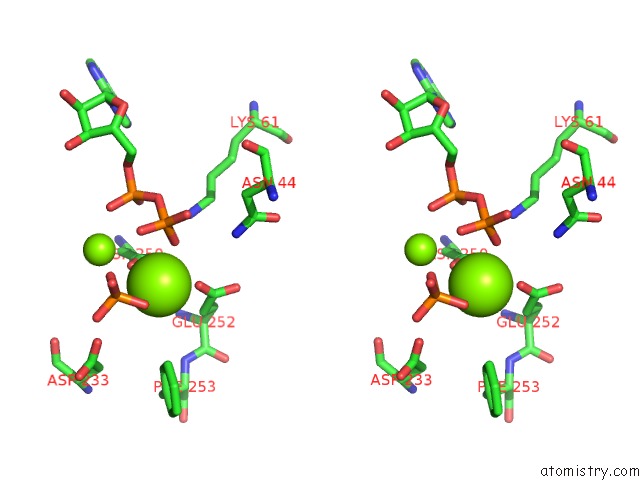
Magnesium binding site 2 out of 4 in 2pup
Go back to
Magnesium binding site 2 out
of 4 in the Structures of 5-Methylthioribose Kinase Reveal Substrate Specificity and Unusual Mode of Nucleotide Binding
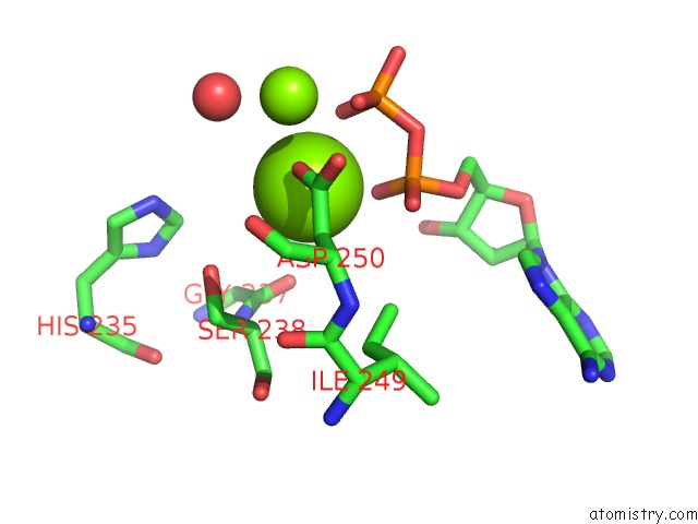
Mono view
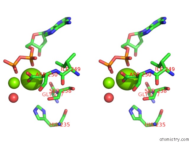
Stereo pair view
Mono view
Stereo pair view
A full contact list of Magnesium with other atoms in the Mg binding
site number 2 of Structures of 5-Methylthioribose Kinase Reveal Substrate Specificity and Unusual Mode of Nucleotide Binding within 5.0Å range:
|
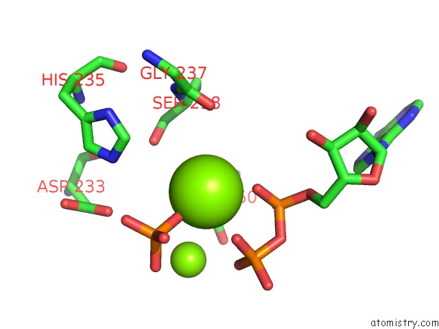
Magnesium binding site 3 out of 4 in 2pup
Go back to
Magnesium binding site 3 out
of 4 in the Structures of 5-Methylthioribose Kinase Reveal Substrate Specificity and Unusual Mode of Nucleotide Binding
Mono view
Stereo pair view
Mono view
Stereo pair view
A full contact list of Magnesium with other atoms in the Mg binding
site number 3 of Structures of 5-Methylthioribose Kinase Reveal Substrate Specificity and Unusual Mode of Nucleotide Binding within 5.0Å range:
|
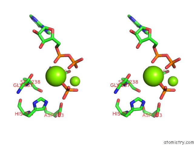
Magnesium binding site 4 out of 4 in 2pup
Go back to
Magnesium binding site 4 out
of 4 in the Structures of 5-Methylthioribose Kinase Reveal Substrate Specificity and Unusual Mode of Nucleotide Binding
Mono view
Stereo pair view
Mono view
Stereo pair view
A full contact list of Magnesium with other atoms in the Mg binding
site number 4 of Structures of 5-Methylthioribose Kinase Reveal Substrate Specificity and Unusual Mode of Nucleotide Binding within 5.0Å range:
|
Reference:
S.-Y.Ku,
P.Yip,
K.A.Cornell,
M.K.Riscoe,
J.-B.Behr,
G.Guillerm,
P.L.Howell.
Structures of 5-Methylthioribose Kinase Reveal Substrate Specificity and Unusual Mode of Nucleotide Binding J.Biol.Chem. V. 282 22195 2007.
ISSN: ISSN 0021-9258
PubMed: 17522047
DOI: 10.1074/JBC.M611045200
Page generated: Wed Aug 14 02:18:17 2024
ISSN: ISSN 0021-9258
PubMed: 17522047
DOI: 10.1074/JBC.M611045200
Last articles
Cl in 7VZZCl in 7VYQ
Cl in 7VZN
Cl in 7VZQ
Cl in 7VYP
Cl in 7VWN
Cl in 7VYO
Cl in 7VVT
Cl in 7VUK
Cl in 7VS9