Magnesium »
PDB 2q0f-2q9p »
2q5l »
Magnesium in PDB 2q5l: X-Ray Structure of Phenylpyruvate Decarboxylase in Complex with 2-(1- Hydroxyethyl)-3-Deaza-Thdp
Enzymatic activity of X-Ray Structure of Phenylpyruvate Decarboxylase in Complex with 2-(1- Hydroxyethyl)-3-Deaza-Thdp
All present enzymatic activity of X-Ray Structure of Phenylpyruvate Decarboxylase in Complex with 2-(1- Hydroxyethyl)-3-Deaza-Thdp:
4.1.1.43;
4.1.1.43;
Protein crystallography data
The structure of X-Ray Structure of Phenylpyruvate Decarboxylase in Complex with 2-(1- Hydroxyethyl)-3-Deaza-Thdp, PDB code: 2q5l
was solved by
W.Versees,
S.Spaepen,
M.D.Wood,
F.J.Leeper,
J.Vanderleyden,
J.Steyaert,
with X-Ray Crystallography technique. A brief refinement statistics is given in the table below:
| Resolution Low / High (Å) | 32.50 / 1.85 |
| Space group | C 2 2 21 |
| Cell size a, b, c (Å), α, β, γ (°) | 99.978, 179.050, 120.858, 90.00, 90.00, 90.00 |
| R / Rfree (%) | 17 / 20.4 |
Other elements in 2q5l:
The structure of X-Ray Structure of Phenylpyruvate Decarboxylase in Complex with 2-(1- Hydroxyethyl)-3-Deaza-Thdp also contains other interesting chemical elements:
| Chlorine | (Cl) | 2 atoms |
Magnesium Binding Sites:
The binding sites of Magnesium atom in the X-Ray Structure of Phenylpyruvate Decarboxylase in Complex with 2-(1- Hydroxyethyl)-3-Deaza-Thdp
(pdb code 2q5l). This binding sites where shown within
5.0 Angstroms radius around Magnesium atom.
In total 2 binding sites of Magnesium where determined in the X-Ray Structure of Phenylpyruvate Decarboxylase in Complex with 2-(1- Hydroxyethyl)-3-Deaza-Thdp, PDB code: 2q5l:
Jump to Magnesium binding site number: 1; 2;
In total 2 binding sites of Magnesium where determined in the X-Ray Structure of Phenylpyruvate Decarboxylase in Complex with 2-(1- Hydroxyethyl)-3-Deaza-Thdp, PDB code: 2q5l:
Jump to Magnesium binding site number: 1; 2;
Magnesium binding site 1 out of 2 in 2q5l
Go back to
Magnesium binding site 1 out
of 2 in the X-Ray Structure of Phenylpyruvate Decarboxylase in Complex with 2-(1- Hydroxyethyl)-3-Deaza-Thdp
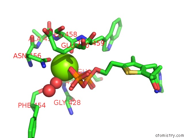
Mono view
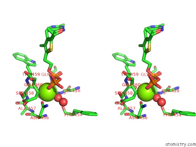
Stereo pair view
Mono view
Stereo pair view
A full contact list of Magnesium with other atoms in the Mg binding
site number 1 of X-Ray Structure of Phenylpyruvate Decarboxylase in Complex with 2-(1- Hydroxyethyl)-3-Deaza-Thdp within 5.0Å range:
|
Magnesium binding site 2 out of 2 in 2q5l
Go back to
Magnesium binding site 2 out
of 2 in the X-Ray Structure of Phenylpyruvate Decarboxylase in Complex with 2-(1- Hydroxyethyl)-3-Deaza-Thdp
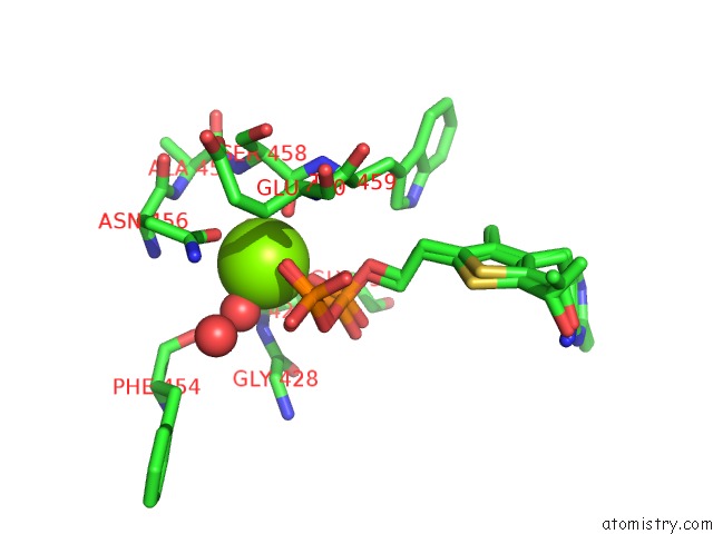
Mono view
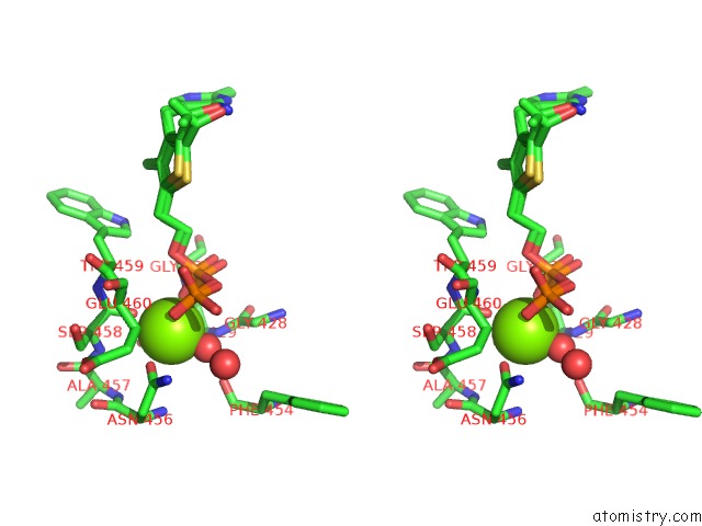
Stereo pair view
Mono view
Stereo pair view
A full contact list of Magnesium with other atoms in the Mg binding
site number 2 of X-Ray Structure of Phenylpyruvate Decarboxylase in Complex with 2-(1- Hydroxyethyl)-3-Deaza-Thdp within 5.0Å range:
|
Reference:
W.Versees,
S.Spaepen,
M.D.Wood,
F.J.Leeper,
J.Vanderleyden,
J.Steyaert.
Molecular Mechanism of Allosteric Substrate Activation in A Thiamine Diphosphate-Dependent Decarboxylase. J.Biol.Chem. V. 282 35269 2007.
ISSN: ISSN 0021-9258
PubMed: 17905741
DOI: 10.1074/JBC.M706048200
Page generated: Wed Aug 14 02:36:19 2024
ISSN: ISSN 0021-9258
PubMed: 17905741
DOI: 10.1074/JBC.M706048200
Last articles
Cl in 8DZ7Cl in 8DYZ
Cl in 8DZ0
Cl in 8DYH
Cl in 8DYD
Cl in 8DXU
Cl in 8DY5
Cl in 8DVC
Cl in 8DUY
Cl in 8DX8