Magnesium »
PDB 2q0f-2q9p »
2q7t »
Magnesium in PDB 2q7t: Crystal Structure of the F Plasmid Trai Relaxase Domain with the Scissile Thymidine Base
Protein crystallography data
The structure of Crystal Structure of the F Plasmid Trai Relaxase Domain with the Scissile Thymidine Base, PDB code: 2q7t
was solved by
S.A.Lujan,
M.R.Redinbo,
with X-Ray Crystallography technique. A brief refinement statistics is given in the table below:
| Resolution Low / High (Å) | 44.13 / 2.42 |
| Space group | P 21 21 21 |
| Cell size a, b, c (Å), α, β, γ (°) | 44.888, 88.259, 127.293, 90.00, 90.00, 90.00 |
| R / Rfree (%) | 21.1 / 27 |
Magnesium Binding Sites:
The binding sites of Magnesium atom in the Crystal Structure of the F Plasmid Trai Relaxase Domain with the Scissile Thymidine Base
(pdb code 2q7t). This binding sites where shown within
5.0 Angstroms radius around Magnesium atom.
In total 2 binding sites of Magnesium where determined in the Crystal Structure of the F Plasmid Trai Relaxase Domain with the Scissile Thymidine Base, PDB code: 2q7t:
Jump to Magnesium binding site number: 1; 2;
In total 2 binding sites of Magnesium where determined in the Crystal Structure of the F Plasmid Trai Relaxase Domain with the Scissile Thymidine Base, PDB code: 2q7t:
Jump to Magnesium binding site number: 1; 2;
Magnesium binding site 1 out of 2 in 2q7t
Go back to
Magnesium binding site 1 out
of 2 in the Crystal Structure of the F Plasmid Trai Relaxase Domain with the Scissile Thymidine Base
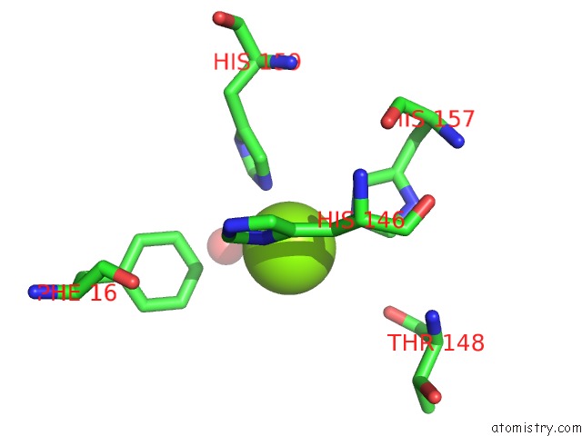
Mono view
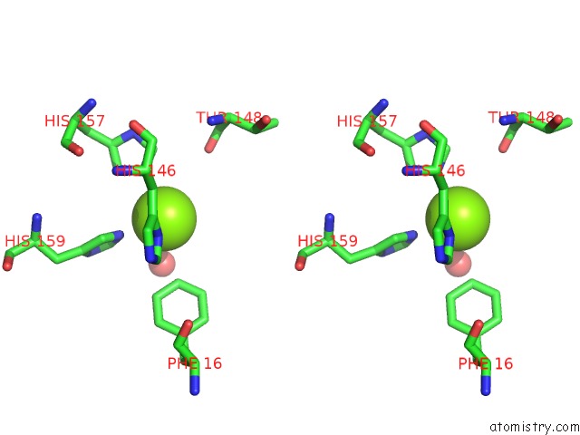
Stereo pair view
Mono view
Stereo pair view
A full contact list of Magnesium with other atoms in the Mg binding
site number 1 of Crystal Structure of the F Plasmid Trai Relaxase Domain with the Scissile Thymidine Base within 5.0Å range:
|
Magnesium binding site 2 out of 2 in 2q7t
Go back to
Magnesium binding site 2 out
of 2 in the Crystal Structure of the F Plasmid Trai Relaxase Domain with the Scissile Thymidine Base
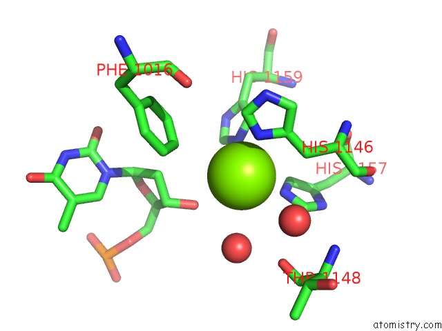
Mono view
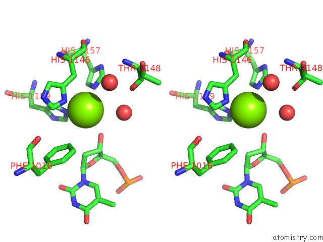
Stereo pair view
Mono view
Stereo pair view
A full contact list of Magnesium with other atoms in the Mg binding
site number 2 of Crystal Structure of the F Plasmid Trai Relaxase Domain with the Scissile Thymidine Base within 5.0Å range:
|
Reference:
S.A.Lujan,
L.M.Guogas,
H.Ragonese,
S.W.Matson,
M.R.Redinbo.
Disrupting Antibiotic Resistance Propagation By Inhibiting the Conjugative Dna Relaxase. Proc.Natl.Acad.Sci.Usa V. 104 12282 2007.
ISSN: ISSN 0027-8424
PubMed: 17630285
DOI: 10.1073/PNAS.0702760104
Page generated: Wed Aug 14 02:38:13 2024
ISSN: ISSN 0027-8424
PubMed: 17630285
DOI: 10.1073/PNAS.0702760104
Last articles
Zn in 9MJ5Zn in 9HNW
Zn in 9G0L
Zn in 9FNE
Zn in 9DZN
Zn in 9E0I
Zn in 9D32
Zn in 9DAK
Zn in 8ZXC
Zn in 8ZUF