Magnesium »
PDB 2q9y-2qrf »
2qjn »
Magnesium in PDB 2qjn: Crystal Structure of D-Mannonate Dehydratase From Novosphingobium Aromaticivorans Complexed with Mg and 2-Keto-3-Deoxy-D-Gluconate
Protein crystallography data
The structure of Crystal Structure of D-Mannonate Dehydratase From Novosphingobium Aromaticivorans Complexed with Mg and 2-Keto-3-Deoxy-D-Gluconate, PDB code: 2qjn
was solved by
A.A.Fedorov,
E.V.Fedorov,
J.F.Rakus,
J.E.Vick,
J.A.Gerlt,
S.C.Almo,
with X-Ray Crystallography technique. A brief refinement statistics is given in the table below:
| Resolution Low / High (Å) | 24.89 / 2.00 |
| Space group | C 2 2 21 |
| Cell size a, b, c (Å), α, β, γ (°) | 116.269, 165.241, 167.039, 90.00, 90.00, 90.00 |
| R / Rfree (%) | 16.2 / 19.1 |
Magnesium Binding Sites:
The binding sites of Magnesium atom in the Crystal Structure of D-Mannonate Dehydratase From Novosphingobium Aromaticivorans Complexed with Mg and 2-Keto-3-Deoxy-D-Gluconate
(pdb code 2qjn). This binding sites where shown within
5.0 Angstroms radius around Magnesium atom.
In total 4 binding sites of Magnesium where determined in the Crystal Structure of D-Mannonate Dehydratase From Novosphingobium Aromaticivorans Complexed with Mg and 2-Keto-3-Deoxy-D-Gluconate, PDB code: 2qjn:
Jump to Magnesium binding site number: 1; 2; 3; 4;
In total 4 binding sites of Magnesium where determined in the Crystal Structure of D-Mannonate Dehydratase From Novosphingobium Aromaticivorans Complexed with Mg and 2-Keto-3-Deoxy-D-Gluconate, PDB code: 2qjn:
Jump to Magnesium binding site number: 1; 2; 3; 4;
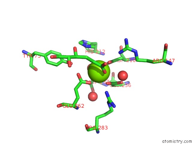
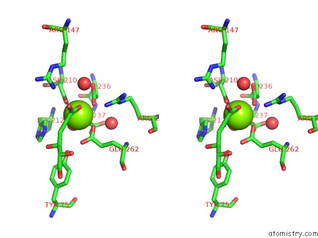
Magnesium binding site 1 out of 4 in 2qjn
Go back to
Magnesium binding site 1 out
of 4 in the Crystal Structure of D-Mannonate Dehydratase From Novosphingobium Aromaticivorans Complexed with Mg and 2-Keto-3-Deoxy-D-Gluconate
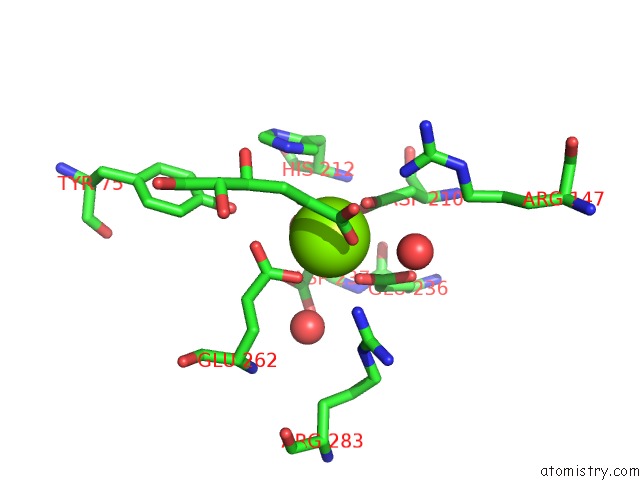
Mono view
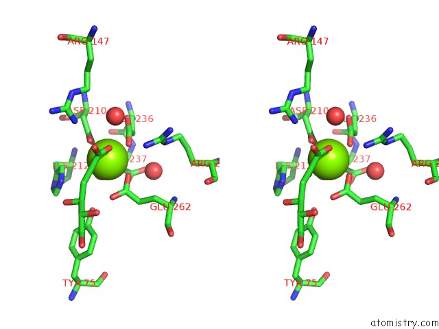
Stereo pair view
Mono view
Stereo pair view
A full contact list of Magnesium with other atoms in the Mg binding
site number 1 of Crystal Structure of D-Mannonate Dehydratase From Novosphingobium Aromaticivorans Complexed with Mg and 2-Keto-3-Deoxy-D-Gluconate within 5.0Å range:
|
Magnesium binding site 2 out of 4 in 2qjn
Go back to
Magnesium binding site 2 out
of 4 in the Crystal Structure of D-Mannonate Dehydratase From Novosphingobium Aromaticivorans Complexed with Mg and 2-Keto-3-Deoxy-D-Gluconate
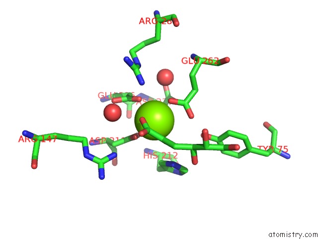
Mono view
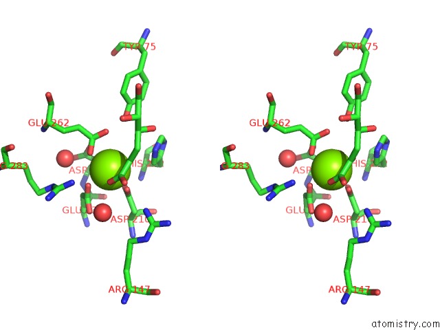
Stereo pair view
Mono view
Stereo pair view
A full contact list of Magnesium with other atoms in the Mg binding
site number 2 of Crystal Structure of D-Mannonate Dehydratase From Novosphingobium Aromaticivorans Complexed with Mg and 2-Keto-3-Deoxy-D-Gluconate within 5.0Å range:
|
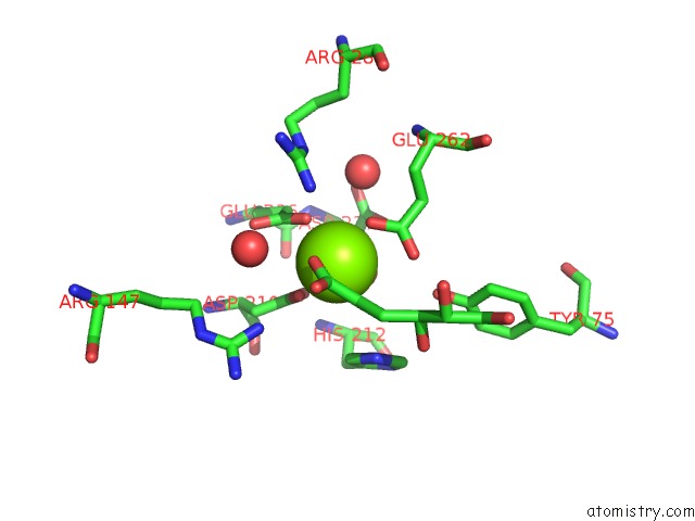
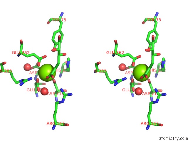
Magnesium binding site 3 out of 4 in 2qjn
Go back to
Magnesium binding site 3 out
of 4 in the Crystal Structure of D-Mannonate Dehydratase From Novosphingobium Aromaticivorans Complexed with Mg and 2-Keto-3-Deoxy-D-Gluconate
Mono view
Stereo pair view
Mono view
Stereo pair view
A full contact list of Magnesium with other atoms in the Mg binding
site number 3 of Crystal Structure of D-Mannonate Dehydratase From Novosphingobium Aromaticivorans Complexed with Mg and 2-Keto-3-Deoxy-D-Gluconate within 5.0Å range:
|
Magnesium binding site 4 out of 4 in 2qjn
Go back to
Magnesium binding site 4 out
of 4 in the Crystal Structure of D-Mannonate Dehydratase From Novosphingobium Aromaticivorans Complexed with Mg and 2-Keto-3-Deoxy-D-Gluconate
Mono view
Stereo pair view
Mono view
Stereo pair view
A full contact list of Magnesium with other atoms in the Mg binding
site number 4 of Crystal Structure of D-Mannonate Dehydratase From Novosphingobium Aromaticivorans Complexed with Mg and 2-Keto-3-Deoxy-D-Gluconate within 5.0Å range:
|
Reference:
J.F.Rakus,
A.A.Fedorov,
E.V.Fedorov,
M.E.Glasner,
J.E.Vick,
P.C.Babbitt,
S.C.Almo,
J.A.Gerlt.
Evolution of Enzymatic Activities in the Enolase Superfamily: D-Mannonate Dehydratase From Novosphingobium Aromaticivorans. Biochemistry V. 46 12896 2007.
ISSN: ISSN 0006-2960
PubMed: 17944491
DOI: 10.1021/BI701703W
Page generated: Wed Aug 14 02:45:54 2024
ISSN: ISSN 0006-2960
PubMed: 17944491
DOI: 10.1021/BI701703W
Last articles
F in 4HY6F in 4HXN
F in 4HT2
F in 4HU1
F in 4HVS
F in 4HW7
F in 4HUA
F in 4HU9
F in 4HQJ
F in 4HT3