Magnesium »
PDB 2q9y-2qrf »
2qq6 »
Magnesium in PDB 2qq6: Crystal Structure of Mandelate Racemase/Muconate Lactonizing Enzyme-Like Protein From Rubrobacter Xylanophilus Dsm 9941
Protein crystallography data
The structure of Crystal Structure of Mandelate Racemase/Muconate Lactonizing Enzyme-Like Protein From Rubrobacter Xylanophilus Dsm 9941, PDB code: 2qq6
was solved by
S.Eswaramoorthy,
M.Madegowda,
S.K.Burley,
S.Swaminathan,
Newyork Sgx Research Center For Structural Genomics (Nysgxrc),
with X-Ray Crystallography technique. A brief refinement statistics is given in the table below:
| Resolution Low / High (Å) | 50.00 / 2.90 |
| Space group | I 41 2 2 |
| Cell size a, b, c (Å), α, β, γ (°) | 184.369, 184.369, 118.108, 90.00, 90.00, 90.00 |
| R / Rfree (%) | 22.2 / 28.2 |
Magnesium Binding Sites:
The binding sites of Magnesium atom in the Crystal Structure of Mandelate Racemase/Muconate Lactonizing Enzyme-Like Protein From Rubrobacter Xylanophilus Dsm 9941
(pdb code 2qq6). This binding sites where shown within
5.0 Angstroms radius around Magnesium atom.
In total 2 binding sites of Magnesium where determined in the Crystal Structure of Mandelate Racemase/Muconate Lactonizing Enzyme-Like Protein From Rubrobacter Xylanophilus Dsm 9941, PDB code: 2qq6:
Jump to Magnesium binding site number: 1; 2;
In total 2 binding sites of Magnesium where determined in the Crystal Structure of Mandelate Racemase/Muconate Lactonizing Enzyme-Like Protein From Rubrobacter Xylanophilus Dsm 9941, PDB code: 2qq6:
Jump to Magnesium binding site number: 1; 2;
Magnesium binding site 1 out of 2 in 2qq6
Go back to
Magnesium binding site 1 out
of 2 in the Crystal Structure of Mandelate Racemase/Muconate Lactonizing Enzyme-Like Protein From Rubrobacter Xylanophilus Dsm 9941
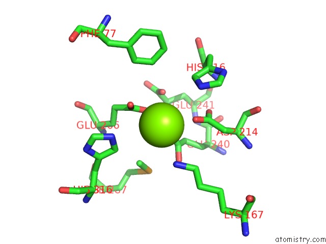
Mono view
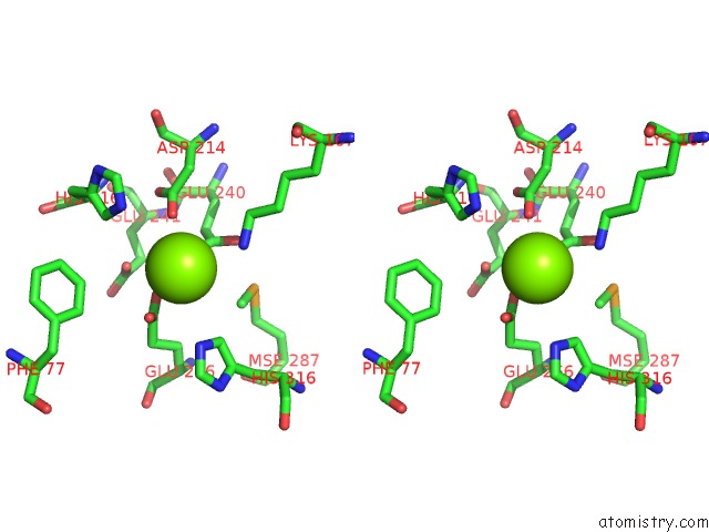
Stereo pair view
Mono view
Stereo pair view
A full contact list of Magnesium with other atoms in the Mg binding
site number 1 of Crystal Structure of Mandelate Racemase/Muconate Lactonizing Enzyme-Like Protein From Rubrobacter Xylanophilus Dsm 9941 within 5.0Å range:
|
Magnesium binding site 2 out of 2 in 2qq6
Go back to
Magnesium binding site 2 out
of 2 in the Crystal Structure of Mandelate Racemase/Muconate Lactonizing Enzyme-Like Protein From Rubrobacter Xylanophilus Dsm 9941
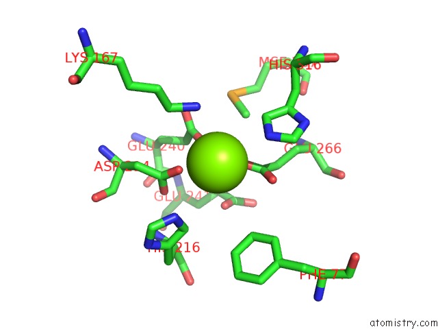
Mono view
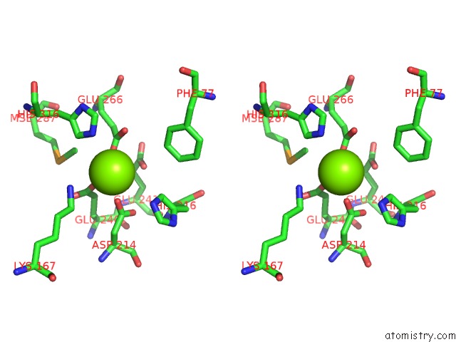
Stereo pair view
Mono view
Stereo pair view
A full contact list of Magnesium with other atoms in the Mg binding
site number 2 of Crystal Structure of Mandelate Racemase/Muconate Lactonizing Enzyme-Like Protein From Rubrobacter Xylanophilus Dsm 9941 within 5.0Å range:
|
Reference:
S.Eswaramoorthy,
M.Madegowda,
S.K.Burley,
S.Swaminathan.
Crystal Structure of Mandelate Racemase/Muconate Lactonizing Enzyme-Like Protein From Rubrobacter Xylanophilus Dsm 9941. To Be Published.
Page generated: Sun Aug 10 13:25:54 2025
Last articles
Mg in 3G5AMg in 3G5S
Mg in 3G58
Mg in 3G4T
Mg in 3G4L
Mg in 3G4K
Mg in 3G4I
Mg in 3G4G
Mg in 3G4F
Mg in 3G45