Magnesium »
PDB 2qrn-2r1y »
2qv7 »
Magnesium in PDB 2qv7: Crystal Structure of Diacylglycerol Kinase Dgkb in Complex with Adp and Mg
Enzymatic activity of Crystal Structure of Diacylglycerol Kinase Dgkb in Complex with Adp and Mg
All present enzymatic activity of Crystal Structure of Diacylglycerol Kinase Dgkb in Complex with Adp and Mg:
2.7.1.107;
2.7.1.107;
Protein crystallography data
The structure of Crystal Structure of Diacylglycerol Kinase Dgkb in Complex with Adp and Mg, PDB code: 2qv7
was solved by
D.J.Miller,
A.Jerga,
C.O.Rock,
S.W.White,
with X-Ray Crystallography technique. A brief refinement statistics is given in the table below:
| Resolution Low / High (Å) | 29.17 / 2.30 |
| Space group | P 42 21 2 |
| Cell size a, b, c (Å), α, β, γ (°) | 123.760, 123.760, 47.680, 90.00, 90.00, 90.00 |
| R / Rfree (%) | 20.6 / 24.6 |
Magnesium Binding Sites:
The binding sites of Magnesium atom in the Crystal Structure of Diacylglycerol Kinase Dgkb in Complex with Adp and Mg
(pdb code 2qv7). This binding sites where shown within
5.0 Angstroms radius around Magnesium atom.
In total only one binding site of Magnesium was determined in the Crystal Structure of Diacylglycerol Kinase Dgkb in Complex with Adp and Mg, PDB code: 2qv7:
In total only one binding site of Magnesium was determined in the Crystal Structure of Diacylglycerol Kinase Dgkb in Complex with Adp and Mg, PDB code: 2qv7:
Magnesium binding site 1 out of 1 in 2qv7
Go back to
Magnesium binding site 1 out
of 1 in the Crystal Structure of Diacylglycerol Kinase Dgkb in Complex with Adp and Mg
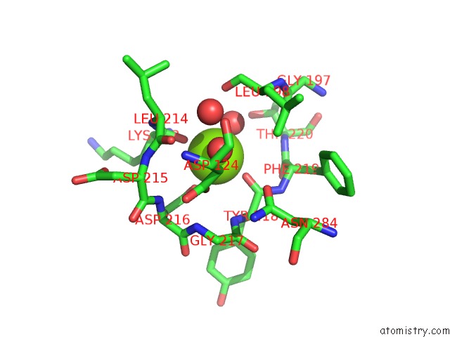
Mono view
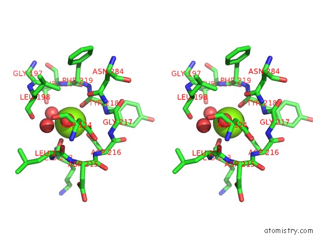
Stereo pair view
Mono view
Stereo pair view
A full contact list of Magnesium with other atoms in the Mg binding
site number 1 of Crystal Structure of Diacylglycerol Kinase Dgkb in Complex with Adp and Mg within 5.0Å range:
|
Reference:
D.J.Miller,
A.Jerga,
C.O.Rock,
S.W.White.
Analysis of the Staphylococcus Aureus Dgkb Structure Reveals A Common Catalytic Mechanism For the Soluble Diacylglycerol Kinases. Structure V. 16 1036 2008.
ISSN: ISSN 0969-2126
PubMed: 18611377
DOI: 10.1016/J.STR.2008.03.019
Page generated: Wed Aug 14 03:10:56 2024
ISSN: ISSN 0969-2126
PubMed: 18611377
DOI: 10.1016/J.STR.2008.03.019
Last articles
Zn in 9JYWZn in 9IR4
Zn in 9IR3
Zn in 9GMX
Zn in 9GMW
Zn in 9JEJ
Zn in 9ERF
Zn in 9ERE
Zn in 9EGV
Zn in 9EGW