Magnesium »
PDB 2qrn-2r1y »
2qvr »
Magnesium in PDB 2qvr: E. Coli Fructose-1,6-Bisphosphatase: Citrate, Fru-2,6-P2, and MG2+ Bound
Enzymatic activity of E. Coli Fructose-1,6-Bisphosphatase: Citrate, Fru-2,6-P2, and MG2+ Bound
All present enzymatic activity of E. Coli Fructose-1,6-Bisphosphatase: Citrate, Fru-2,6-P2, and MG2+ Bound:
3.1.3.11;
3.1.3.11;
Protein crystallography data
The structure of E. Coli Fructose-1,6-Bisphosphatase: Citrate, Fru-2,6-P2, and MG2+ Bound, PDB code: 2qvr
was solved by
J.K.Hines,
H.J.Fromm,
R.B.Honzatko,
with X-Ray Crystallography technique. A brief refinement statistics is given in the table below:
| Resolution Low / High (Å) | 47.44 / 2.18 |
| Space group | I 2 2 2 |
| Cell size a, b, c (Å), α, β, γ (°) | 44.002, 82.251, 174.194, 90.00, 90.00, 90.00 |
| R / Rfree (%) | 19.6 / 24.2 |
Magnesium Binding Sites:
The binding sites of Magnesium atom in the E. Coli Fructose-1,6-Bisphosphatase: Citrate, Fru-2,6-P2, and MG2+ Bound
(pdb code 2qvr). This binding sites where shown within
5.0 Angstroms radius around Magnesium atom.
In total only one binding site of Magnesium was determined in the E. Coli Fructose-1,6-Bisphosphatase: Citrate, Fru-2,6-P2, and MG2+ Bound, PDB code: 2qvr:
In total only one binding site of Magnesium was determined in the E. Coli Fructose-1,6-Bisphosphatase: Citrate, Fru-2,6-P2, and MG2+ Bound, PDB code: 2qvr:
Magnesium binding site 1 out of 1 in 2qvr
Go back to
Magnesium binding site 1 out
of 1 in the E. Coli Fructose-1,6-Bisphosphatase: Citrate, Fru-2,6-P2, and MG2+ Bound
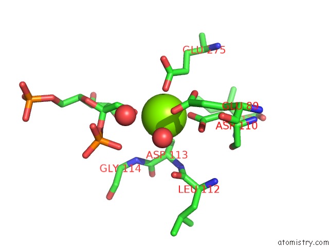
Mono view
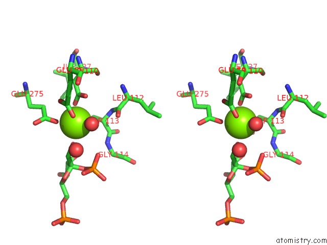
Stereo pair view
Mono view
Stereo pair view
A full contact list of Magnesium with other atoms in the Mg binding
site number 1 of E. Coli Fructose-1,6-Bisphosphatase: Citrate, Fru-2,6-P2, and MG2+ Bound within 5.0Å range:
|
Reference:
J.K.Hines,
X.Chen,
J.C.Nix,
H.J.Fromm,
R.B.Honzatko.
Structures of Mammalian and Bacterial Fructose-1,6-Bisphosphatase Reveal the Basis For Synergism in Amp/Fructose 2,6-Bisphosphate Inhibition. J.Biol.Chem. V. 282 36121 2007.
ISSN: ISSN 0021-9258
PubMed: 17933867
DOI: 10.1074/JBC.M707302200
Page generated: Wed Aug 14 03:11:22 2024
ISSN: ISSN 0021-9258
PubMed: 17933867
DOI: 10.1074/JBC.M707302200
Last articles
Zn in 9JYWZn in 9IR4
Zn in 9IR3
Zn in 9GMX
Zn in 9GMW
Zn in 9JEJ
Zn in 9ERF
Zn in 9ERE
Zn in 9EGV
Zn in 9EGW