Magnesium »
PDB 2r20-2reu »
2r6t »
Magnesium in PDB 2r6t: Structure of A R132K Variant Pduo-Type Atp:Co(I)Rrinoid Adenosyltransferase From Lactobacillus Reuteri Complexed with Atp
Protein crystallography data
The structure of Structure of A R132K Variant Pduo-Type Atp:Co(I)Rrinoid Adenosyltransferase From Lactobacillus Reuteri Complexed with Atp, PDB code: 2r6t
was solved by
M.St Maurice,
P.E.Mera,
J.C.Escalante-Semerena,
I.Rayment,
with X-Ray Crystallography technique. A brief refinement statistics is given in the table below:
| Resolution Low / High (Å) | 30.00 / 2.61 |
| Space group | P 63 |
| Cell size a, b, c (Å), α, β, γ (°) | 64.678, 64.678, 169.197, 90.00, 90.00, 120.00 |
| R / Rfree (%) | 18.9 / 25.9 |
Magnesium Binding Sites:
The binding sites of Magnesium atom in the Structure of A R132K Variant Pduo-Type Atp:Co(I)Rrinoid Adenosyltransferase From Lactobacillus Reuteri Complexed with Atp
(pdb code 2r6t). This binding sites where shown within
5.0 Angstroms radius around Magnesium atom.
In total 4 binding sites of Magnesium where determined in the Structure of A R132K Variant Pduo-Type Atp:Co(I)Rrinoid Adenosyltransferase From Lactobacillus Reuteri Complexed with Atp, PDB code: 2r6t:
Jump to Magnesium binding site number: 1; 2; 3; 4;
In total 4 binding sites of Magnesium where determined in the Structure of A R132K Variant Pduo-Type Atp:Co(I)Rrinoid Adenosyltransferase From Lactobacillus Reuteri Complexed with Atp, PDB code: 2r6t:
Jump to Magnesium binding site number: 1; 2; 3; 4;
Magnesium binding site 1 out of 4 in 2r6t
Go back to
Magnesium binding site 1 out
of 4 in the Structure of A R132K Variant Pduo-Type Atp:Co(I)Rrinoid Adenosyltransferase From Lactobacillus Reuteri Complexed with Atp

Mono view
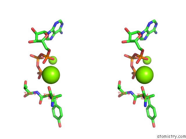
Stereo pair view
Mono view
Stereo pair view
A full contact list of Magnesium with other atoms in the Mg binding
site number 1 of Structure of A R132K Variant Pduo-Type Atp:Co(I)Rrinoid Adenosyltransferase From Lactobacillus Reuteri Complexed with Atp within 5.0Å range:
|
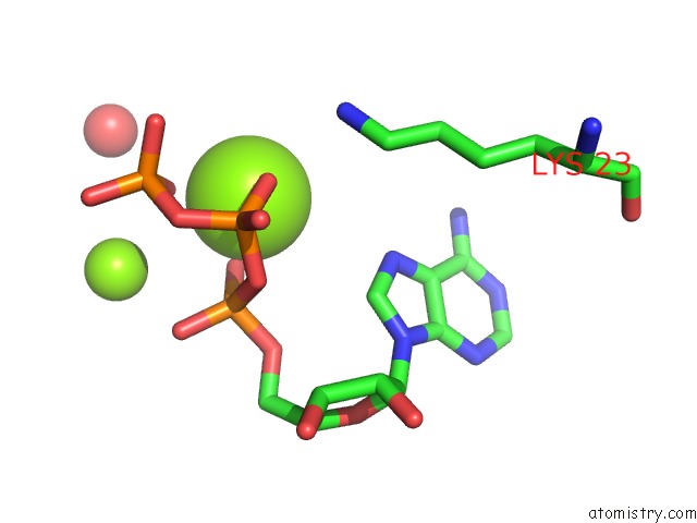
Magnesium binding site 2 out of 4 in 2r6t
Go back to
Magnesium binding site 2 out
of 4 in the Structure of A R132K Variant Pduo-Type Atp:Co(I)Rrinoid Adenosyltransferase From Lactobacillus Reuteri Complexed with Atp

Mono view

Stereo pair view
Mono view
Stereo pair view
A full contact list of Magnesium with other atoms in the Mg binding
site number 2 of Structure of A R132K Variant Pduo-Type Atp:Co(I)Rrinoid Adenosyltransferase From Lactobacillus Reuteri Complexed with Atp within 5.0Å range:
|
Magnesium binding site 3 out of 4 in 2r6t
Go back to
Magnesium binding site 3 out
of 4 in the Structure of A R132K Variant Pduo-Type Atp:Co(I)Rrinoid Adenosyltransferase From Lactobacillus Reuteri Complexed with Atp
Mono view
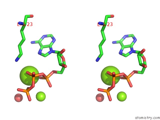
Stereo pair view
Mono view
Stereo pair view
A full contact list of Magnesium with other atoms in the Mg binding
site number 3 of Structure of A R132K Variant Pduo-Type Atp:Co(I)Rrinoid Adenosyltransferase From Lactobacillus Reuteri Complexed with Atp within 5.0Å range:
|
Magnesium binding site 4 out of 4 in 2r6t
Go back to
Magnesium binding site 4 out
of 4 in the Structure of A R132K Variant Pduo-Type Atp:Co(I)Rrinoid Adenosyltransferase From Lactobacillus Reuteri Complexed with Atp
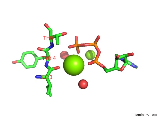
Mono view

Stereo pair view
Mono view
Stereo pair view
A full contact list of Magnesium with other atoms in the Mg binding
site number 4 of Structure of A R132K Variant Pduo-Type Atp:Co(I)Rrinoid Adenosyltransferase From Lactobacillus Reuteri Complexed with Atp within 5.0Å range:
|
Reference:
P.E.Mera,
M.St Maurice,
I.Rayment,
J.C.Escalante-Semerena.
Structural and Functional Analyses of the Human-Type Corrinoid Adenosyltransferase (Pduo) From Lactobacillus Reuteri. Biochemistry V. 46 13829 2007.
ISSN: ISSN 0006-2960
PubMed: 17988155
DOI: 10.1021/BI701622J
Page generated: Wed Aug 14 03:16:44 2024
ISSN: ISSN 0006-2960
PubMed: 17988155
DOI: 10.1021/BI701622J
Last articles
F in 4F2XF in 4EZJ
F in 4EWS
F in 4ELF
F in 4EWQ
F in 4EQU
F in 4EST
F in 4ENH
F in 4EPX
F in 4ENC