Magnesium »
PDB 2r20-2reu »
2r6x »
Magnesium in PDB 2r6x: Structure of A D35N Variant Pduo-Type Atp:Co(I)Rrinoid Adenosyltransferase From Lactobacillus Reuteri Complexed with Atp
Enzymatic activity of Structure of A D35N Variant Pduo-Type Atp:Co(I)Rrinoid Adenosyltransferase From Lactobacillus Reuteri Complexed with Atp
All present enzymatic activity of Structure of A D35N Variant Pduo-Type Atp:Co(I)Rrinoid Adenosyltransferase From Lactobacillus Reuteri Complexed with Atp:
2.5.1.17;
2.5.1.17;
Protein crystallography data
The structure of Structure of A D35N Variant Pduo-Type Atp:Co(I)Rrinoid Adenosyltransferase From Lactobacillus Reuteri Complexed with Atp, PDB code: 2r6x
was solved by
M.St Maurice,
P.E.Mera,
J.C.Escalante-Semerena,
I.Rayment,
with X-Ray Crystallography technique. A brief refinement statistics is given in the table below:
| Resolution Low / High (Å) | 30.00 / 2.61 |
| Space group | P 63 |
| Cell size a, b, c (Å), α, β, γ (°) | 64.772, 64.772, 169.438, 90.00, 90.00, 120.00 |
| R / Rfree (%) | 17.3 / 24.9 |
Magnesium Binding Sites:
The binding sites of Magnesium atom in the Structure of A D35N Variant Pduo-Type Atp:Co(I)Rrinoid Adenosyltransferase From Lactobacillus Reuteri Complexed with Atp
(pdb code 2r6x). This binding sites where shown within
5.0 Angstroms radius around Magnesium atom.
In total 4 binding sites of Magnesium where determined in the Structure of A D35N Variant Pduo-Type Atp:Co(I)Rrinoid Adenosyltransferase From Lactobacillus Reuteri Complexed with Atp, PDB code: 2r6x:
Jump to Magnesium binding site number: 1; 2; 3; 4;
In total 4 binding sites of Magnesium where determined in the Structure of A D35N Variant Pduo-Type Atp:Co(I)Rrinoid Adenosyltransferase From Lactobacillus Reuteri Complexed with Atp, PDB code: 2r6x:
Jump to Magnesium binding site number: 1; 2; 3; 4;
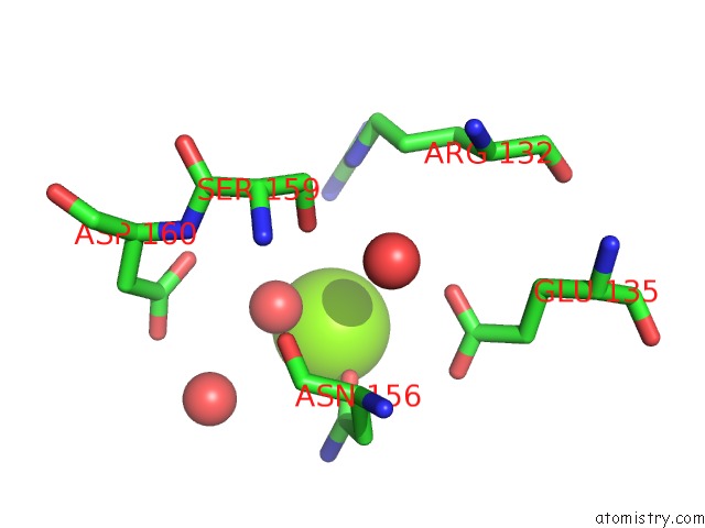
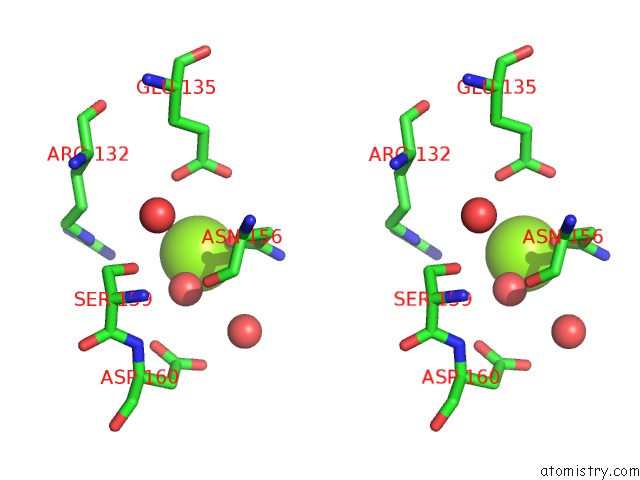
Magnesium binding site 1 out of 4 in 2r6x
Go back to
Magnesium binding site 1 out
of 4 in the Structure of A D35N Variant Pduo-Type Atp:Co(I)Rrinoid Adenosyltransferase From Lactobacillus Reuteri Complexed with Atp
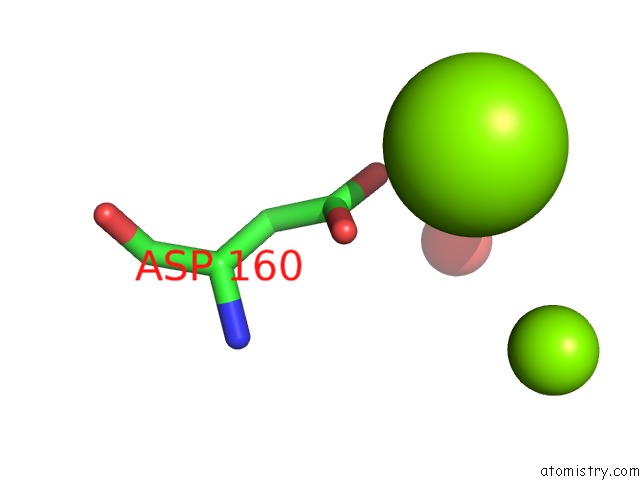
Mono view
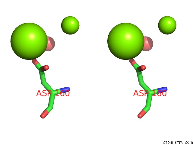
Stereo pair view
Mono view
Stereo pair view
A full contact list of Magnesium with other atoms in the Mg binding
site number 1 of Structure of A D35N Variant Pduo-Type Atp:Co(I)Rrinoid Adenosyltransferase From Lactobacillus Reuteri Complexed with Atp within 5.0Å range:
|
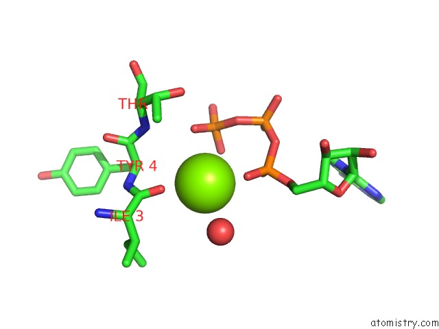
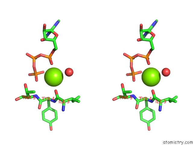
Magnesium binding site 2 out of 4 in 2r6x
Go back to
Magnesium binding site 2 out
of 4 in the Structure of A D35N Variant Pduo-Type Atp:Co(I)Rrinoid Adenosyltransferase From Lactobacillus Reuteri Complexed with Atp
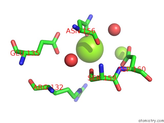
Mono view
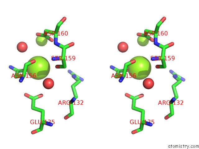
Stereo pair view
Mono view
Stereo pair view
A full contact list of Magnesium with other atoms in the Mg binding
site number 2 of Structure of A D35N Variant Pduo-Type Atp:Co(I)Rrinoid Adenosyltransferase From Lactobacillus Reuteri Complexed with Atp within 5.0Å range:
|
Magnesium binding site 3 out of 4 in 2r6x
Go back to
Magnesium binding site 3 out
of 4 in the Structure of A D35N Variant Pduo-Type Atp:Co(I)Rrinoid Adenosyltransferase From Lactobacillus Reuteri Complexed with Atp
Mono view
Stereo pair view
Mono view
Stereo pair view
A full contact list of Magnesium with other atoms in the Mg binding
site number 3 of Structure of A D35N Variant Pduo-Type Atp:Co(I)Rrinoid Adenosyltransferase From Lactobacillus Reuteri Complexed with Atp within 5.0Å range:
|
Magnesium binding site 4 out of 4 in 2r6x
Go back to
Magnesium binding site 4 out
of 4 in the Structure of A D35N Variant Pduo-Type Atp:Co(I)Rrinoid Adenosyltransferase From Lactobacillus Reuteri Complexed with Atp
Mono view
Stereo pair view
Mono view
Stereo pair view
A full contact list of Magnesium with other atoms in the Mg binding
site number 4 of Structure of A D35N Variant Pduo-Type Atp:Co(I)Rrinoid Adenosyltransferase From Lactobacillus Reuteri Complexed with Atp within 5.0Å range:
|
Reference:
P.E.Mera,
M.St Maurice,
I.Rayment,
J.C.Escalante-Semerena.
Structural and Functional Analyses of the Human-Type Corrinoid Adenosyltransferase (Pduo) From Lactobacillus Reuteri. Biochemistry V. 46 13829 2007.
ISSN: ISSN 0006-2960
PubMed: 17988155
DOI: 10.1021/BI701622J
Page generated: Wed Aug 14 03:16:44 2024
ISSN: ISSN 0006-2960
PubMed: 17988155
DOI: 10.1021/BI701622J
Last articles
Fe in 2YXOFe in 2YRS
Fe in 2YXC
Fe in 2YNM
Fe in 2YVJ
Fe in 2YP1
Fe in 2YU2
Fe in 2YU1
Fe in 2YQB
Fe in 2YOO