Magnesium »
PDB 2r20-2reu »
2r72 »
Magnesium in PDB 2r72: Crystal Structure of Infectious Bursal Disease Virus VP1 Polymerase, Incubated with MG2+ Ion.
Protein crystallography data
The structure of Crystal Structure of Infectious Bursal Disease Virus VP1 Polymerase, Incubated with MG2+ Ion., PDB code: 2r72
was solved by
D.Garriga,
A.Navarro,
J.Querol-Audi,
F.Abaitua,
J.F.Rodriguez,
N.Verdaguer,
with X-Ray Crystallography technique. A brief refinement statistics is given in the table below:
| Resolution Low / High (Å) | 20.00 / 3.15 |
| Space group | P 61 2 2 |
| Cell size a, b, c (Å), α, β, γ (°) | 122.838, 122.838, 354.255, 90.00, 90.00, 120.00 |
| R / Rfree (%) | 20.9 / 23.5 |
Magnesium Binding Sites:
The binding sites of Magnesium atom in the Crystal Structure of Infectious Bursal Disease Virus VP1 Polymerase, Incubated with MG2+ Ion.
(pdb code 2r72). This binding sites where shown within
5.0 Angstroms radius around Magnesium atom.
In total 3 binding sites of Magnesium where determined in the Crystal Structure of Infectious Bursal Disease Virus VP1 Polymerase, Incubated with MG2+ Ion., PDB code: 2r72:
Jump to Magnesium binding site number: 1; 2; 3;
In total 3 binding sites of Magnesium where determined in the Crystal Structure of Infectious Bursal Disease Virus VP1 Polymerase, Incubated with MG2+ Ion., PDB code: 2r72:
Jump to Magnesium binding site number: 1; 2; 3;
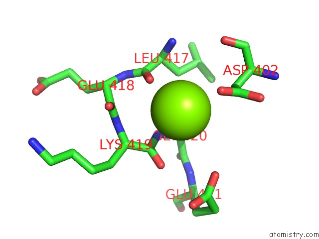
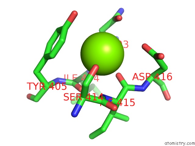
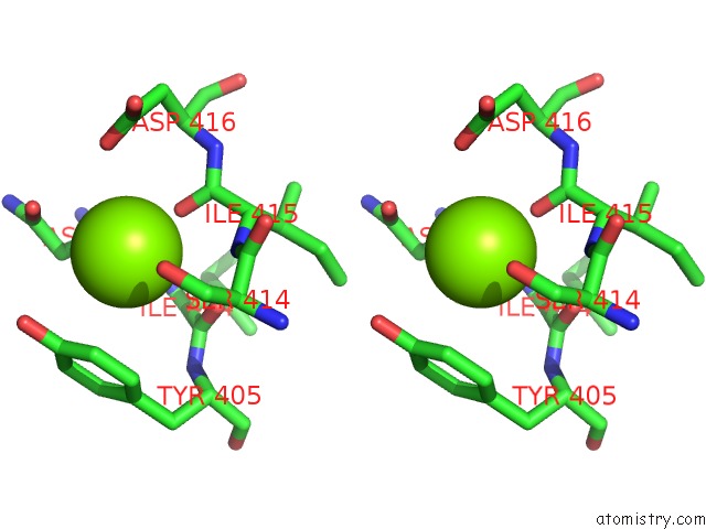
Magnesium binding site 1 out of 3 in 2r72
Go back to
Magnesium binding site 1 out
of 3 in the Crystal Structure of Infectious Bursal Disease Virus VP1 Polymerase, Incubated with MG2+ Ion.
Mono view
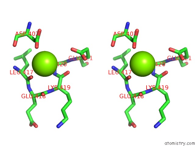
Stereo pair view
Mono view
Stereo pair view
A full contact list of Magnesium with other atoms in the Mg binding
site number 1 of Crystal Structure of Infectious Bursal Disease Virus VP1 Polymerase, Incubated with MG2+ Ion. within 5.0Å range:
|
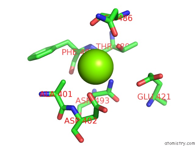
Magnesium binding site 2 out of 3 in 2r72
Go back to
Magnesium binding site 2 out
of 3 in the Crystal Structure of Infectious Bursal Disease Virus VP1 Polymerase, Incubated with MG2+ Ion.
Mono view
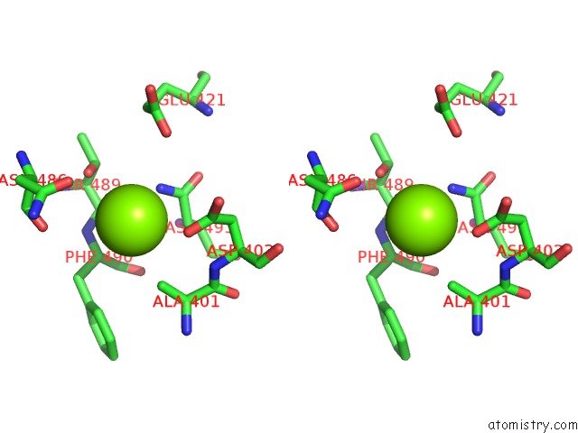
Stereo pair view
Mono view
Stereo pair view
A full contact list of Magnesium with other atoms in the Mg binding
site number 2 of Crystal Structure of Infectious Bursal Disease Virus VP1 Polymerase, Incubated with MG2+ Ion. within 5.0Å range:
|
Magnesium binding site 3 out of 3 in 2r72
Go back to
Magnesium binding site 3 out
of 3 in the Crystal Structure of Infectious Bursal Disease Virus VP1 Polymerase, Incubated with MG2+ Ion.
Mono view
Stereo pair view
Mono view
Stereo pair view
A full contact list of Magnesium with other atoms in the Mg binding
site number 3 of Crystal Structure of Infectious Bursal Disease Virus VP1 Polymerase, Incubated with MG2+ Ion. within 5.0Å range:
|
Reference:
D.Garriga,
A.Navarro,
J.Querol-Audi,
F.Abaitua,
J.F.Rodriguez,
N.Verdaguer.
Activation Mechanism of A Noncanonical Rna-Dependent Rna Polymerase. Proc.Natl.Acad.Sci.Usa V. 104 20540 2007.
ISSN: ISSN 0027-8424
PubMed: 18077388
DOI: 10.1073/PNAS.0704447104
Page generated: Wed Aug 14 03:17:01 2024
ISSN: ISSN 0027-8424
PubMed: 18077388
DOI: 10.1073/PNAS.0704447104
Last articles
Cl in 6ARCCl in 6AQY
Cl in 6AQE
Cl in 6ALJ
Cl in 6AQ4
Cl in 6AOM
Cl in 6APQ
Cl in 6AP6
Cl in 6AOL
Cl in 6AOK