Magnesium »
PDB 2uxj-2v7y »
2v1w »
Magnesium in PDB 2v1w: Crystal Structure of Human Lim Protein Ril (PDLIM4) Pdz Domain Bound to the C-Terminal Peptide of Human Alpha-Actinin-1
Protein crystallography data
The structure of Crystal Structure of Human Lim Protein Ril (PDLIM4) Pdz Domain Bound to the C-Terminal Peptide of Human Alpha-Actinin-1, PDB code: 2v1w
was solved by
M.Soundararajan,
L.Shrestha,
A.C.W.Pike,
E.Salah,
N.Burgess-Brown,
J.Elkins,
C.Umeano,
E.Ugochukwu,
F.Von Delft,
C.H.Arrowsmith,
A.Edwards,
J.Weigelt,
M.Sundstrom,
D.Doyle,
with X-Ray Crystallography technique. A brief refinement statistics is given in the table below:
| Resolution Low / High (Å) | 61.55 / 1.90 |
| Space group | P 4 21 2 |
| Cell size a, b, c (Å), α, β, γ (°) | 87.029, 87.029, 53.917, 90.00, 90.00, 90.00 |
| R / Rfree (%) | 17.3 / 21.1 |
Magnesium Binding Sites:
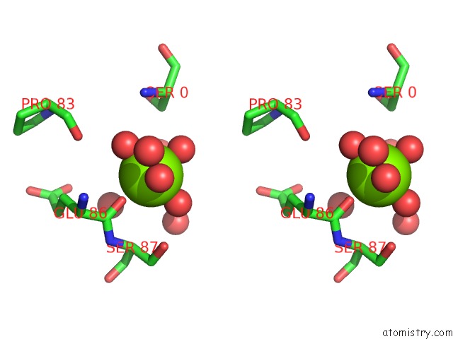
The binding sites of Magnesium atom in the Crystal Structure of Human Lim Protein Ril (PDLIM4) Pdz Domain Bound to the C-Terminal Peptide of Human Alpha-Actinin-1
(pdb code 2v1w). This binding sites where shown within
5.0 Angstroms radius around Magnesium atom.
In total only one binding site of Magnesium was determined in the Crystal Structure of Human Lim Protein Ril (PDLIM4) Pdz Domain Bound to the C-Terminal Peptide of Human Alpha-Actinin-1, PDB code: 2v1w:
In total only one binding site of Magnesium was determined in the Crystal Structure of Human Lim Protein Ril (PDLIM4) Pdz Domain Bound to the C-Terminal Peptide of Human Alpha-Actinin-1, PDB code: 2v1w:
Magnesium binding site 1 out of 1 in 2v1w
Go back to
Magnesium binding site 1 out
of 1 in the Crystal Structure of Human Lim Protein Ril (PDLIM4) Pdz Domain Bound to the C-Terminal Peptide of Human Alpha-Actinin-1

Mono view
Stereo pair view
Mono view
Stereo pair view
A full contact list of Magnesium with other atoms in the Mg binding
site number 1 of Crystal Structure of Human Lim Protein Ril (PDLIM4) Pdz Domain Bound to the C-Terminal Peptide of Human Alpha-Actinin-1 within 5.0Å range:
|
Reference:
J.M.Elkins,
C.Gileadi,
L.Shrestha,
C.Phillips,
J.Wang,
J.R.C.Muniz,
D.A.Doyle.
Unusual Binding Interactions in Pdz Domain Crystal Structures Help Explain Binding Mechanisms. Protein Sci. V. 19 731 2010.
ISSN: ISSN 0961-8368
PubMed: 20120020
DOI: 10.1002/PRO.349
Page generated: Wed Aug 14 04:57:23 2024
ISSN: ISSN 0961-8368
PubMed: 20120020
DOI: 10.1002/PRO.349
Last articles
Zn in 9J0NZn in 9J0O
Zn in 9J0P
Zn in 9FJX
Zn in 9EKB
Zn in 9C0F
Zn in 9CAH
Zn in 9CH0
Zn in 9CH3
Zn in 9CH1