Magnesium »
PDB 2uxj-2v7y »
2v52 »
Magnesium in PDB 2v52: Structure of Mal-RPEL2 Complexed to G-Actin
Protein crystallography data
The structure of Structure of Mal-RPEL2 Complexed to G-Actin, PDB code: 2v52
was solved by
S.Mouilleron,
S.Guettler,
C.A.Langer,
R.Treisman,
N.Q.Mcdonald,
with X-Ray Crystallography technique. A brief refinement statistics is given in the table below:
| Resolution Low / High (Å) | 29.35 / 1.45 |
| Space group | P 21 21 21 |
| Cell size a, b, c (Å), α, β, γ (°) | 54.750, 55.440, 138.390, 90.00, 90.00, 90.00 |
| R / Rfree (%) | 14.7 / 18.8 |
Magnesium Binding Sites:
The binding sites of Magnesium atom in the Structure of Mal-RPEL2 Complexed to G-Actin
(pdb code 2v52). This binding sites where shown within
5.0 Angstroms radius around Magnesium atom.
In total only one binding site of Magnesium was determined in the Structure of Mal-RPEL2 Complexed to G-Actin, PDB code: 2v52:
In total only one binding site of Magnesium was determined in the Structure of Mal-RPEL2 Complexed to G-Actin, PDB code: 2v52:
Magnesium binding site 1 out of 1 in 2v52
Go back to
Magnesium binding site 1 out
of 1 in the Structure of Mal-RPEL2 Complexed to G-Actin
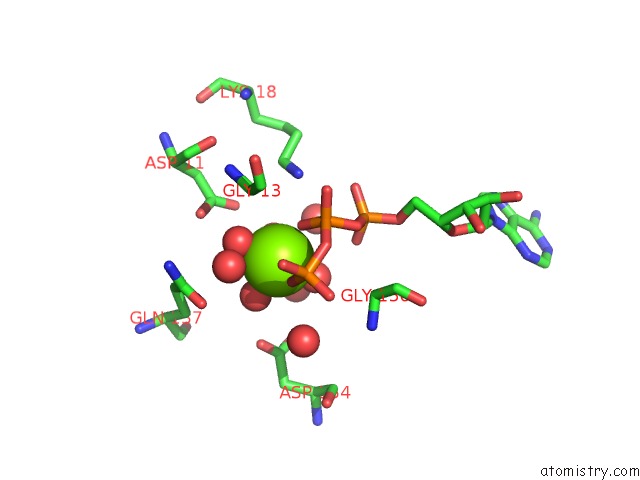
Mono view
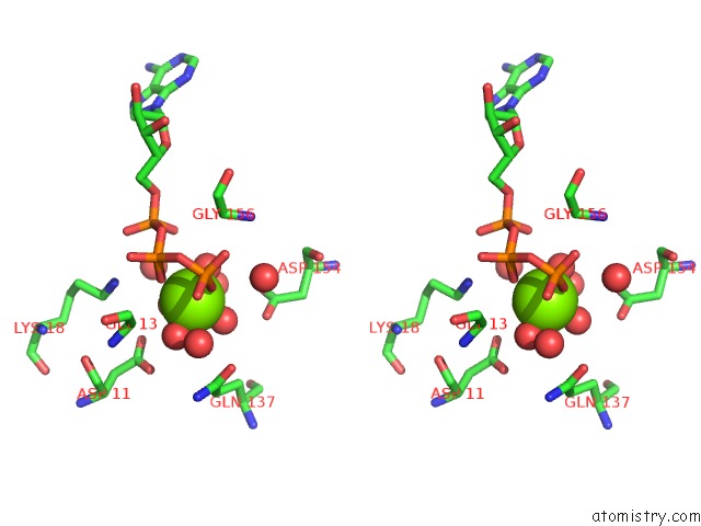
Stereo pair view
Mono view
Stereo pair view
A full contact list of Magnesium with other atoms in the Mg binding
site number 1 of Structure of Mal-RPEL2 Complexed to G-Actin within 5.0Å range:
|
Reference:
S.Mouilleron,
S.Guettler,
C.A.Langer,
R.Treisman,
N.Q.Mcdonald.
Molecular Basis For G-Actin Binding to Rpel Motifs From the Serum Response Factor Coactivator Mal. Embo J. V. 27 3198 2008.
ISSN: ESSN 1460-2075
PubMed: 19008859
DOI: 10.1038/EMBOJ.2008.235
Page generated: Wed Aug 14 04:58:45 2024
ISSN: ESSN 1460-2075
PubMed: 19008859
DOI: 10.1038/EMBOJ.2008.235
Last articles
Zn in 9MJ5Zn in 9HNW
Zn in 9G0L
Zn in 9FNE
Zn in 9DZN
Zn in 9E0I
Zn in 9D32
Zn in 9DAK
Zn in 8ZXC
Zn in 8ZUF