Magnesium »
PDB 2v9j-2vfj »
2vdo »
Magnesium in PDB 2vdo: Integrin ALPHAIIBBETA3 Headpiece Bound to Fibrinogen Gamma Chain Peptide, Hhlggakqagdv
Protein crystallography data
The structure of Integrin ALPHAIIBBETA3 Headpiece Bound to Fibrinogen Gamma Chain Peptide, Hhlggakqagdv, PDB code: 2vdo
was solved by
T.A.Springer,
J.Zhu,
T.Xiao,
with X-Ray Crystallography technique. A brief refinement statistics is given in the table below:
| Resolution Low / High (Å) | 46.13 / 2.51 |
| Space group | P 32 2 1 |
| Cell size a, b, c (Å), α, β, γ (°) | 148.331, 148.331, 176.567, 90.00, 90.00, 120.00 |
| R / Rfree (%) | 14.8 / 19 |
Other elements in 2vdo:
The structure of Integrin ALPHAIIBBETA3 Headpiece Bound to Fibrinogen Gamma Chain Peptide, Hhlggakqagdv also contains other interesting chemical elements:
| Calcium | (Ca) | 6 atoms |
Magnesium Binding Sites:
The binding sites of Magnesium atom in the Integrin ALPHAIIBBETA3 Headpiece Bound to Fibrinogen Gamma Chain Peptide, Hhlggakqagdv
(pdb code 2vdo). This binding sites where shown within
5.0 Angstroms radius around Magnesium atom.
In total only one binding site of Magnesium was determined in the Integrin ALPHAIIBBETA3 Headpiece Bound to Fibrinogen Gamma Chain Peptide, Hhlggakqagdv, PDB code: 2vdo:
In total only one binding site of Magnesium was determined in the Integrin ALPHAIIBBETA3 Headpiece Bound to Fibrinogen Gamma Chain Peptide, Hhlggakqagdv, PDB code: 2vdo:
Magnesium binding site 1 out of 1 in 2vdo
Go back to
Magnesium binding site 1 out
of 1 in the Integrin ALPHAIIBBETA3 Headpiece Bound to Fibrinogen Gamma Chain Peptide, Hhlggakqagdv
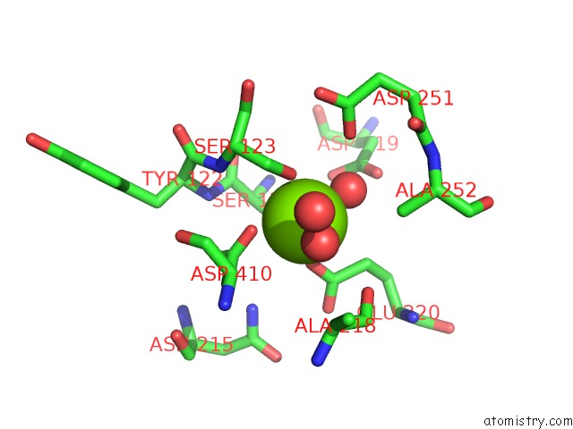
Mono view
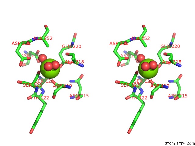
Stereo pair view
Mono view
Stereo pair view
A full contact list of Magnesium with other atoms in the Mg binding
site number 1 of Integrin ALPHAIIBBETA3 Headpiece Bound to Fibrinogen Gamma Chain Peptide, Hhlggakqagdv within 5.0Å range:
|
Reference:
T.A.Springer,
J.Zhu,
T.Xiao.
Structural Basis For Distinctive Recognition of Fibrinogen Gammac Peptide By the Platelet Integrin ALPHAIIBBETA3. J.Cell Biol. V. 182 791 2008.
ISSN: ISSN 0021-9525
PubMed: 18710925
DOI: 10.1083/JCB.200801146
Page generated: Wed Aug 14 05:09:43 2024
ISSN: ISSN 0021-9525
PubMed: 18710925
DOI: 10.1083/JCB.200801146
Last articles
Zn in 9J0NZn in 9J0O
Zn in 9J0P
Zn in 9FJX
Zn in 9EKB
Zn in 9C0F
Zn in 9CAH
Zn in 9CH0
Zn in 9CH3
Zn in 9CH1