Magnesium »
PDB 2v9j-2vfj »
2vfj »
Magnesium in PDB 2vfj: Structure of the A20 Ovarian Tumour (Otu) Domain
Enzymatic activity of Structure of the A20 Ovarian Tumour (Otu) Domain
All present enzymatic activity of Structure of the A20 Ovarian Tumour (Otu) Domain:
3.4.19.12;
3.4.19.12;
Protein crystallography data
The structure of Structure of the A20 Ovarian Tumour (Otu) Domain, PDB code: 2vfj
was solved by
D.Komander,
D.Barford,
with X-Ray Crystallography technique. A brief refinement statistics is given in the table below:
| Resolution Low / High (Å) | 50.286 / 3.20 |
| Space group | P 1 21 1 |
| Cell size a, b, c (Å), α, β, γ (°) | 84.964, 83.023, 164.940, 90.00, 98.08, 90.00 |
| R / Rfree (%) | 20.44 / 24.29 |
Magnesium Binding Sites:
The binding sites of Magnesium atom in the Structure of the A20 Ovarian Tumour (Otu) Domain
(pdb code 2vfj). This binding sites where shown within
5.0 Angstroms radius around Magnesium atom.
In total only one binding site of Magnesium was determined in the Structure of the A20 Ovarian Tumour (Otu) Domain, PDB code: 2vfj:
In total only one binding site of Magnesium was determined in the Structure of the A20 Ovarian Tumour (Otu) Domain, PDB code: 2vfj:
Magnesium binding site 1 out of 1 in 2vfj
Go back to
Magnesium binding site 1 out
of 1 in the Structure of the A20 Ovarian Tumour (Otu) Domain
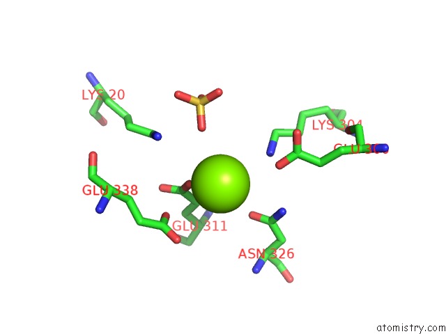
Mono view
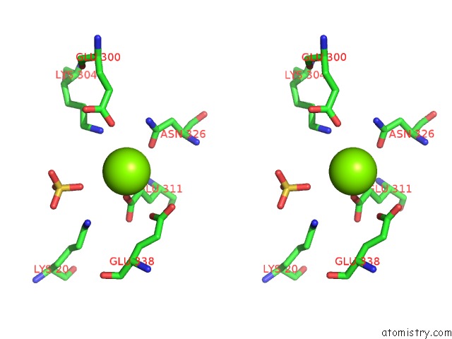
Stereo pair view
Mono view
Stereo pair view
A full contact list of Magnesium with other atoms in the Mg binding
site number 1 of Structure of the A20 Ovarian Tumour (Otu) Domain within 5.0Å range:
|
Reference:
D.Komander,
D.Barford.
Structure of the A20 Otu Domain and Mechanistic Insights Into Deubiquitination Biochem.J. V. 409 77 2008.
ISSN: ISSN 0264-6021
PubMed: 17961127
DOI: 10.1042/BJ20071399
Page generated: Wed Aug 14 05:11:29 2024
ISSN: ISSN 0264-6021
PubMed: 17961127
DOI: 10.1042/BJ20071399
Last articles
Zn in 9MJ5Zn in 9HNW
Zn in 9G0L
Zn in 9FNE
Zn in 9DZN
Zn in 9E0I
Zn in 9D32
Zn in 9DAK
Zn in 8ZXC
Zn in 8ZUF