Magnesium »
PDB 2vfx-2vsc »
2vhx »
Magnesium in PDB 2vhx: Crystal Structure of the Ternary Complex of L-Alanine Dehydrogenase From Mycobacterium Tuberculosis with Nad+ and Pyruvate
Enzymatic activity of Crystal Structure of the Ternary Complex of L-Alanine Dehydrogenase From Mycobacterium Tuberculosis with Nad+ and Pyruvate
All present enzymatic activity of Crystal Structure of the Ternary Complex of L-Alanine Dehydrogenase From Mycobacterium Tuberculosis with Nad+ and Pyruvate:
1.4.1.1;
1.4.1.1;
Protein crystallography data
The structure of Crystal Structure of the Ternary Complex of L-Alanine Dehydrogenase From Mycobacterium Tuberculosis with Nad+ and Pyruvate, PDB code: 2vhx
was solved by
D.Agren,
G.Schneider,
with X-Ray Crystallography technique. A brief refinement statistics is given in the table below:
| Resolution Low / High (Å) | 40.00 / 2.0 |
| Space group | P 21 21 2 |
| Cell size a, b, c (Å), α, β, γ (°) | 176.247, 171.767, 98.254, 90.00, 90.00, 90.00 |
| R / Rfree (%) | 17.2 / 21 |
Magnesium Binding Sites:
The binding sites of Magnesium atom in the Crystal Structure of the Ternary Complex of L-Alanine Dehydrogenase From Mycobacterium Tuberculosis with Nad+ and Pyruvate
(pdb code 2vhx). This binding sites where shown within
5.0 Angstroms radius around Magnesium atom.
In total 3 binding sites of Magnesium where determined in the Crystal Structure of the Ternary Complex of L-Alanine Dehydrogenase From Mycobacterium Tuberculosis with Nad+ and Pyruvate, PDB code: 2vhx:
Jump to Magnesium binding site number: 1; 2; 3;
In total 3 binding sites of Magnesium where determined in the Crystal Structure of the Ternary Complex of L-Alanine Dehydrogenase From Mycobacterium Tuberculosis with Nad+ and Pyruvate, PDB code: 2vhx:
Jump to Magnesium binding site number: 1; 2; 3;
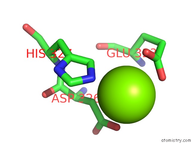
Magnesium binding site 1 out of 3 in 2vhx
Go back to
Magnesium binding site 1 out
of 3 in the Crystal Structure of the Ternary Complex of L-Alanine Dehydrogenase From Mycobacterium Tuberculosis with Nad+ and Pyruvate

Mono view
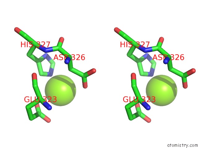
Stereo pair view
Mono view
Stereo pair view
A full contact list of Magnesium with other atoms in the Mg binding
site number 1 of Crystal Structure of the Ternary Complex of L-Alanine Dehydrogenase From Mycobacterium Tuberculosis with Nad+ and Pyruvate within 5.0Å range:
|
Magnesium binding site 2 out of 3 in 2vhx
Go back to
Magnesium binding site 2 out
of 3 in the Crystal Structure of the Ternary Complex of L-Alanine Dehydrogenase From Mycobacterium Tuberculosis with Nad+ and Pyruvate

Mono view

Stereo pair view
Mono view
Stereo pair view
A full contact list of Magnesium with other atoms in the Mg binding
site number 2 of Crystal Structure of the Ternary Complex of L-Alanine Dehydrogenase From Mycobacterium Tuberculosis with Nad+ and Pyruvate within 5.0Å range:
|
Magnesium binding site 3 out of 3 in 2vhx
Go back to
Magnesium binding site 3 out
of 3 in the Crystal Structure of the Ternary Complex of L-Alanine Dehydrogenase From Mycobacterium Tuberculosis with Nad+ and Pyruvate
Mono view

Stereo pair view
Mono view
Stereo pair view
A full contact list of Magnesium with other atoms in the Mg binding
site number 3 of Crystal Structure of the Ternary Complex of L-Alanine Dehydrogenase From Mycobacterium Tuberculosis with Nad+ and Pyruvate within 5.0Å range:
|
Reference:
D.Agren,
M.Stehr,
C.L.Berthold,
S.Kapoor,
W.Oehlmann,
M.Singh,
G.Schneider.
Three-Dimensional Structures of Apo- and Holo-L- Alanine Dehydrogenase From Mycobacterium Tuberculosis Reveal Conformational Changes Upon Coenzyme Binding. J.Mol.Biol. V. 377 1161 2008.
ISSN: ISSN 0022-2836
PubMed: 18304579
DOI: 10.1016/J.JMB.2008.01.091
Page generated: Wed Aug 14 05:13:51 2024
ISSN: ISSN 0022-2836
PubMed: 18304579
DOI: 10.1016/J.JMB.2008.01.091
Last articles
Zn in 9MJ5Zn in 9HNW
Zn in 9G0L
Zn in 9FNE
Zn in 9DZN
Zn in 9E0I
Zn in 9D32
Zn in 9DAK
Zn in 8ZXC
Zn in 8ZUF