Magnesium »
PDB 2vfx-2vsc »
2vn2 »
Magnesium in PDB 2vn2: Crystal Structure of the N-Terminal Domain of Dnad Protein From Geobacillus Kaustophilus HTA426
Protein crystallography data
The structure of Crystal Structure of the N-Terminal Domain of Dnad Protein From Geobacillus Kaustophilus HTA426, PDB code: 2vn2
was solved by
C.-Y.Huang,
Y.-W.Chang,
W.-T.Chen,
Y.-J.Sun,
C.-D.Hsiao,
with X-Ray Crystallography technique. A brief refinement statistics is given in the table below:
| Resolution Low / High (Å) | 27.49 / 2.3 |
| Space group | F 2 2 2 |
| Cell size a, b, c (Å), α, β, γ (°) | 116.514, 124.707, 157.230, 90.00, 90.00, 90.00 |
| R / Rfree (%) | 21.7 / 22.3 |
Magnesium Binding Sites:
The binding sites of Magnesium atom in the Crystal Structure of the N-Terminal Domain of Dnad Protein From Geobacillus Kaustophilus HTA426
(pdb code 2vn2). This binding sites where shown within
5.0 Angstroms radius around Magnesium atom.
In total 4 binding sites of Magnesium where determined in the Crystal Structure of the N-Terminal Domain of Dnad Protein From Geobacillus Kaustophilus HTA426, PDB code: 2vn2:
Jump to Magnesium binding site number: 1; 2; 3; 4;
In total 4 binding sites of Magnesium where determined in the Crystal Structure of the N-Terminal Domain of Dnad Protein From Geobacillus Kaustophilus HTA426, PDB code: 2vn2:
Jump to Magnesium binding site number: 1; 2; 3; 4;
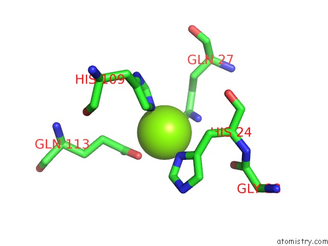
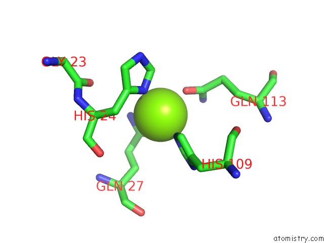
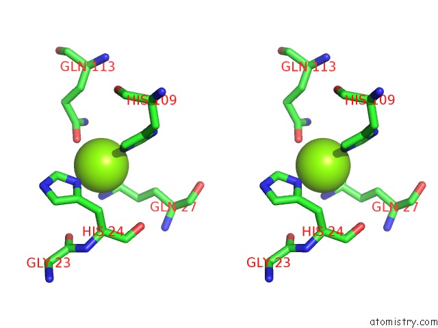
Magnesium binding site 1 out of 4 in 2vn2
Go back to
Magnesium binding site 1 out
of 4 in the Crystal Structure of the N-Terminal Domain of Dnad Protein From Geobacillus Kaustophilus HTA426
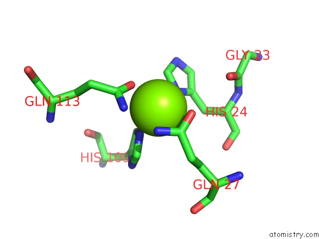
Mono view
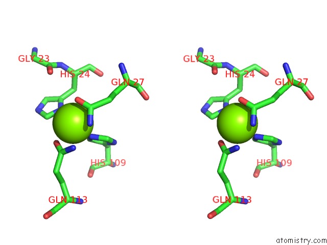
Stereo pair view
Mono view
Stereo pair view
A full contact list of Magnesium with other atoms in the Mg binding
site number 1 of Crystal Structure of the N-Terminal Domain of Dnad Protein From Geobacillus Kaustophilus HTA426 within 5.0Å range:
|
Magnesium binding site 2 out of 4 in 2vn2
Go back to
Magnesium binding site 2 out
of 4 in the Crystal Structure of the N-Terminal Domain of Dnad Protein From Geobacillus Kaustophilus HTA426
Mono view
Stereo pair view
Mono view
Stereo pair view
A full contact list of Magnesium with other atoms in the Mg binding
site number 2 of Crystal Structure of the N-Terminal Domain of Dnad Protein From Geobacillus Kaustophilus HTA426 within 5.0Å range:
|
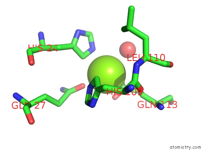
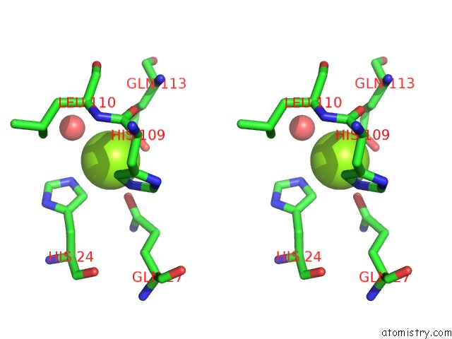
Magnesium binding site 3 out of 4 in 2vn2
Go back to
Magnesium binding site 3 out
of 4 in the Crystal Structure of the N-Terminal Domain of Dnad Protein From Geobacillus Kaustophilus HTA426
Mono view

Stereo pair view
Mono view
Stereo pair view
A full contact list of Magnesium with other atoms in the Mg binding
site number 3 of Crystal Structure of the N-Terminal Domain of Dnad Protein From Geobacillus Kaustophilus HTA426 within 5.0Å range:
|
Magnesium binding site 4 out of 4 in 2vn2
Go back to
Magnesium binding site 4 out
of 4 in the Crystal Structure of the N-Terminal Domain of Dnad Protein From Geobacillus Kaustophilus HTA426
Mono view
Stereo pair view
Mono view
Stereo pair view
A full contact list of Magnesium with other atoms in the Mg binding
site number 4 of Crystal Structure of the N-Terminal Domain of Dnad Protein From Geobacillus Kaustophilus HTA426 within 5.0Å range:
|
Reference:
C.-Y.Huang,
Y.-W.Chang,
W.-T.Chen.
Crystal Structure of the N-Terminal Domain of Geobacillus Kaustophilus HTA426 Dnad Protein. Biochem.Biophys.Res.Commun. V. 375 220 2008.
ISSN: ISSN 0006-291X
PubMed: 18703019
DOI: 10.1016/J.BBRC.2008.07.160
Page generated: Wed Aug 14 05:16:57 2024
ISSN: ISSN 0006-291X
PubMed: 18703019
DOI: 10.1016/J.BBRC.2008.07.160
Last articles
Zn in 9MJ5Zn in 9HNW
Zn in 9G0L
Zn in 9FNE
Zn in 9DZN
Zn in 9E0I
Zn in 9D32
Zn in 9DAK
Zn in 8ZXC
Zn in 8ZUF