Magnesium »
PDB 2vfx-2vsc »
2vpq »
Magnesium in PDB 2vpq: Crystal Structure of Biotin Carboxylase From S. Aureus Complexed with Amppnp
Enzymatic activity of Crystal Structure of Biotin Carboxylase From S. Aureus Complexed with Amppnp
All present enzymatic activity of Crystal Structure of Biotin Carboxylase From S. Aureus Complexed with Amppnp:
6.3.4.14;
6.3.4.14;
Protein crystallography data
The structure of Crystal Structure of Biotin Carboxylase From S. Aureus Complexed with Amppnp, PDB code: 2vpq
was solved by
I.Mochalkin,
with X-Ray Crystallography technique. A brief refinement statistics is given in the table below:
| Resolution Low / High (Å) | 20.00 / 2.1 |
| Space group | P 1 21 1 |
| Cell size a, b, c (Å), α, β, γ (°) | 78.247, 63.318, 105.142, 90.00, 103.82, 90.00 |
| R / Rfree (%) | 21.5 / 28.4 |
Other elements in 2vpq:
The structure of Crystal Structure of Biotin Carboxylase From S. Aureus Complexed with Amppnp also contains other interesting chemical elements:
| Chlorine | (Cl) | 2 atoms |
Magnesium Binding Sites:
The binding sites of Magnesium atom in the Crystal Structure of Biotin Carboxylase From S. Aureus Complexed with Amppnp
(pdb code 2vpq). This binding sites where shown within
5.0 Angstroms radius around Magnesium atom.
In total 4 binding sites of Magnesium where determined in the Crystal Structure of Biotin Carboxylase From S. Aureus Complexed with Amppnp, PDB code: 2vpq:
Jump to Magnesium binding site number: 1; 2; 3; 4;
In total 4 binding sites of Magnesium where determined in the Crystal Structure of Biotin Carboxylase From S. Aureus Complexed with Amppnp, PDB code: 2vpq:
Jump to Magnesium binding site number: 1; 2; 3; 4;
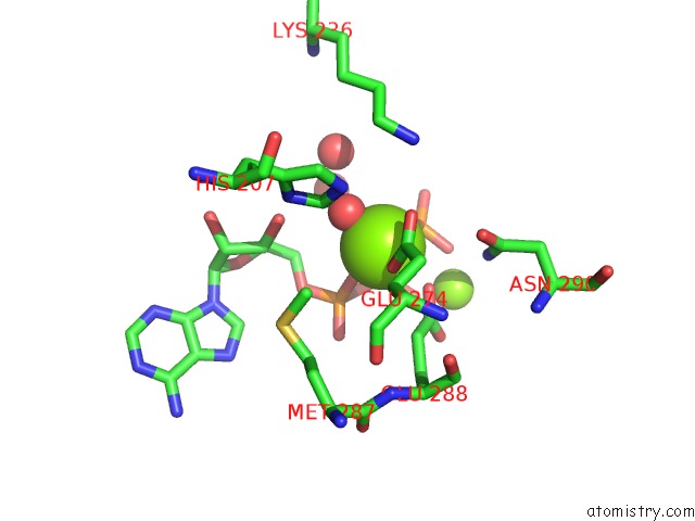
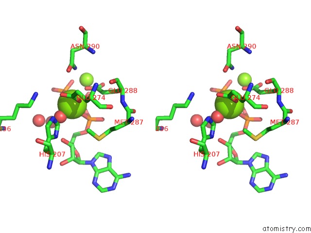
Magnesium binding site 1 out of 4 in 2vpq
Go back to
Magnesium binding site 1 out
of 4 in the Crystal Structure of Biotin Carboxylase From S. Aureus Complexed with Amppnp
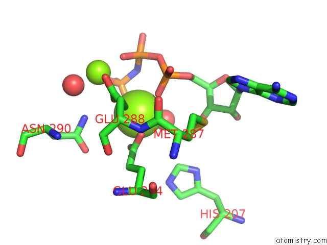
Mono view
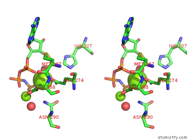
Stereo pair view
Mono view
Stereo pair view
A full contact list of Magnesium with other atoms in the Mg binding
site number 1 of Crystal Structure of Biotin Carboxylase From S. Aureus Complexed with Amppnp within 5.0Å range:
|
Magnesium binding site 2 out of 4 in 2vpq
Go back to
Magnesium binding site 2 out
of 4 in the Crystal Structure of Biotin Carboxylase From S. Aureus Complexed with Amppnp
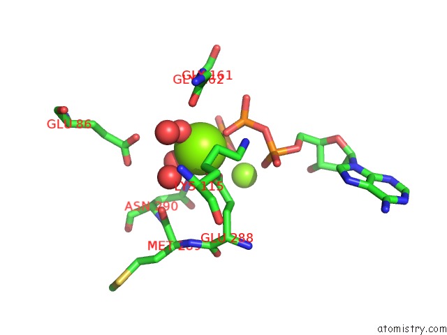
Mono view
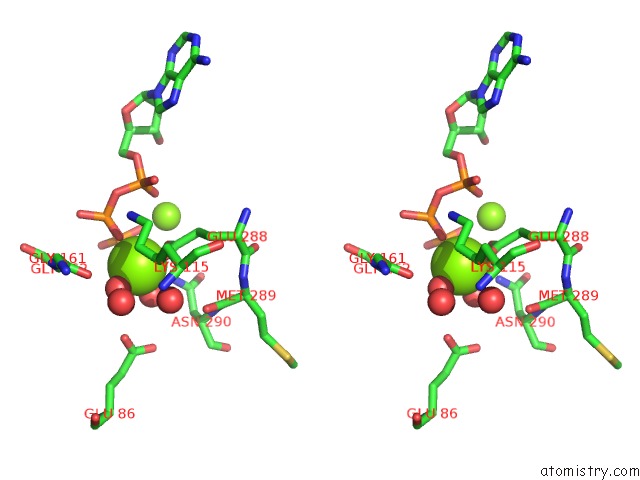
Stereo pair view
Mono view
Stereo pair view
A full contact list of Magnesium with other atoms in the Mg binding
site number 2 of Crystal Structure of Biotin Carboxylase From S. Aureus Complexed with Amppnp within 5.0Å range:
|
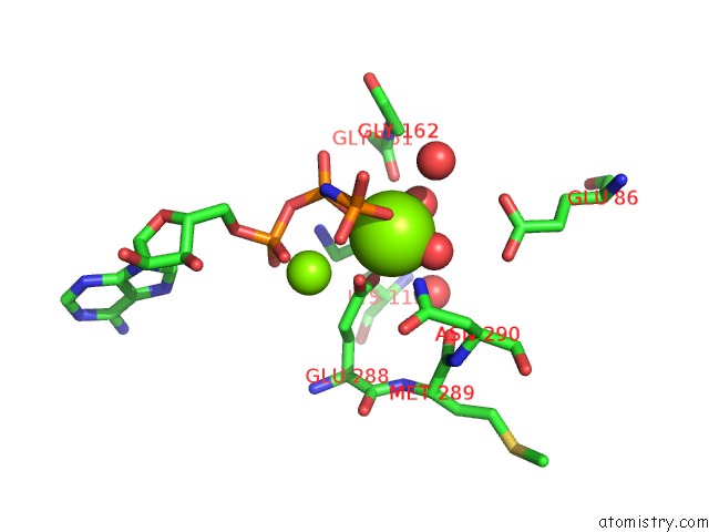
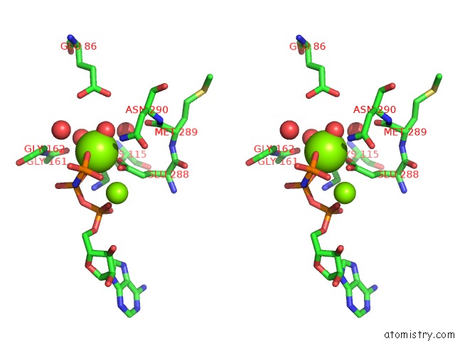
Magnesium binding site 3 out of 4 in 2vpq
Go back to
Magnesium binding site 3 out
of 4 in the Crystal Structure of Biotin Carboxylase From S. Aureus Complexed with Amppnp
Mono view
Stereo pair view
Mono view
Stereo pair view
A full contact list of Magnesium with other atoms in the Mg binding
site number 3 of Crystal Structure of Biotin Carboxylase From S. Aureus Complexed with Amppnp within 5.0Å range:
|
Magnesium binding site 4 out of 4 in 2vpq
Go back to
Magnesium binding site 4 out
of 4 in the Crystal Structure of Biotin Carboxylase From S. Aureus Complexed with Amppnp
Mono view
Stereo pair view
Mono view
Stereo pair view
A full contact list of Magnesium with other atoms in the Mg binding
site number 4 of Crystal Structure of Biotin Carboxylase From S. Aureus Complexed with Amppnp within 5.0Å range:
|
Reference:
I.Mochalkin,
J.R.Miller,
A.Evdokimov,
S.Lightle,
C.Yan,
C.K.Stover,
G.L.Waldrop.
Structural Evidence For Substrate-Induced Synergism and Half-Sites Reactivity in Biotin Carboxylase. Protein Sci. V. 17 1706 2008.
ISSN: ISSN 0961-8368
PubMed: 18725455
DOI: 10.1110/PS.035584.108
Page generated: Wed Aug 14 05:18:01 2024
ISSN: ISSN 0961-8368
PubMed: 18725455
DOI: 10.1110/PS.035584.108
Last articles
Zn in 9MJ5Zn in 9HNW
Zn in 9G0L
Zn in 9FNE
Zn in 9DZN
Zn in 9E0I
Zn in 9D32
Zn in 9DAK
Zn in 8ZXC
Zn in 8ZUF