Magnesium »
PDB 2vfx-2vsc »
2vqd »
Magnesium in PDB 2vqd: Crystal Structure of Biotin Carboxylase From Pseudomonas Aeruginosa Complexed with Ampcp
Enzymatic activity of Crystal Structure of Biotin Carboxylase From Pseudomonas Aeruginosa Complexed with Ampcp
All present enzymatic activity of Crystal Structure of Biotin Carboxylase From Pseudomonas Aeruginosa Complexed with Ampcp:
6.3.4.14;
6.3.4.14;
Protein crystallography data
The structure of Crystal Structure of Biotin Carboxylase From Pseudomonas Aeruginosa Complexed with Ampcp, PDB code: 2vqd
was solved by
I.Mochalkin,
with X-Ray Crystallography technique. A brief refinement statistics is given in the table below:
| Resolution Low / High (Å) | 63.37 / 2.41 |
| Space group | P 21 21 2 |
| Cell size a, b, c (Å), α, β, γ (°) | 64.227, 126.786, 49.878, 90.00, 90.00, 90.00 |
| R / Rfree (%) | 17.8 / 25.2 |
Magnesium Binding Sites:
The binding sites of Magnesium atom in the Crystal Structure of Biotin Carboxylase From Pseudomonas Aeruginosa Complexed with Ampcp
(pdb code 2vqd). This binding sites where shown within
5.0 Angstroms radius around Magnesium atom.
In total only one binding site of Magnesium was determined in the Crystal Structure of Biotin Carboxylase From Pseudomonas Aeruginosa Complexed with Ampcp, PDB code: 2vqd:
In total only one binding site of Magnesium was determined in the Crystal Structure of Biotin Carboxylase From Pseudomonas Aeruginosa Complexed with Ampcp, PDB code: 2vqd:
Magnesium binding site 1 out of 1 in 2vqd
Go back to
Magnesium binding site 1 out
of 1 in the Crystal Structure of Biotin Carboxylase From Pseudomonas Aeruginosa Complexed with Ampcp
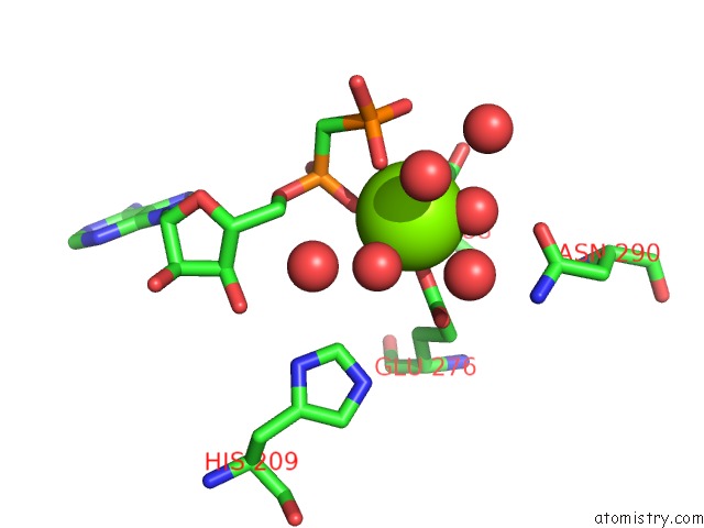
Mono view
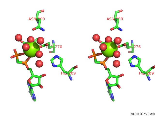
Stereo pair view
Mono view
Stereo pair view
A full contact list of Magnesium with other atoms in the Mg binding
site number 1 of Crystal Structure of Biotin Carboxylase From Pseudomonas Aeruginosa Complexed with Ampcp within 5.0Å range:
|
Reference:
I.Mochalkin,
J.R.Miller,
A.Evdokimov,
S.Lightle,
C.Yan,
C.K.Stover,
G.L.Waldrop.
Structural Evidence For Substrate-Induced Synergism and Half-Sites Reactivity in Biotin Carboxylase. Protein Sci. V. 17 1706 2008.
ISSN: ISSN 0961-8368
PubMed: 18725455
DOI: 10.1110/PS.035584.108
Page generated: Wed Aug 14 05:18:27 2024
ISSN: ISSN 0961-8368
PubMed: 18725455
DOI: 10.1110/PS.035584.108
Last articles
Cl in 5W6FCl in 5W4V
Cl in 5W5J
Cl in 5W5B
Cl in 5W4Q
Cl in 5W5A
Cl in 5W4R
Cl in 5W4P
Cl in 5W4O
Cl in 5W16