Magnesium »
PDB 2vu9-2wb1 »
2vu9 »
Magnesium in PDB 2vu9: Crystal Structure of Botulinum Neurotoxin Serotype A Binding Domain in Complex with GT1B
Enzymatic activity of Crystal Structure of Botulinum Neurotoxin Serotype A Binding Domain in Complex with GT1B
All present enzymatic activity of Crystal Structure of Botulinum Neurotoxin Serotype A Binding Domain in Complex with GT1B:
3.4.24.69;
3.4.24.69;
Protein crystallography data
The structure of Crystal Structure of Botulinum Neurotoxin Serotype A Binding Domain in Complex with GT1B, PDB code: 2vu9
was solved by
P.Stenmark,
J.Dupuy,
R.C.Stevens,
with X-Ray Crystallography technique. A brief refinement statistics is given in the table below:
| Resolution Low / High (Å) | 19.67 / 1.6 |
| Space group | C 2 2 21 |
| Cell size a, b, c (Å), α, β, γ (°) | 72.700, 116.110, 105.520, 90.00, 90.00, 90.00 |
| R / Rfree (%) | 16.1 / 18.6 |
Magnesium Binding Sites:
The binding sites of Magnesium atom in the Crystal Structure of Botulinum Neurotoxin Serotype A Binding Domain in Complex with GT1B
(pdb code 2vu9). This binding sites where shown within
5.0 Angstroms radius around Magnesium atom.
In total only one binding site of Magnesium was determined in the Crystal Structure of Botulinum Neurotoxin Serotype A Binding Domain in Complex with GT1B, PDB code: 2vu9:
In total only one binding site of Magnesium was determined in the Crystal Structure of Botulinum Neurotoxin Serotype A Binding Domain in Complex with GT1B, PDB code: 2vu9:
Magnesium binding site 1 out of 1 in 2vu9
Go back to
Magnesium binding site 1 out
of 1 in the Crystal Structure of Botulinum Neurotoxin Serotype A Binding Domain in Complex with GT1B
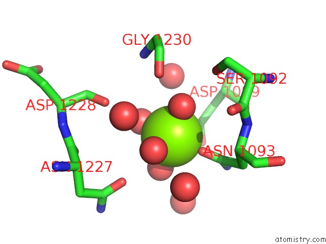
Mono view
Stereo pair view
Mono view
Stereo pair view
A full contact list of Magnesium with other atoms in the Mg binding
site number 1 of Crystal Structure of Botulinum Neurotoxin Serotype A Binding Domain in Complex with GT1B within 5.0Å range:
|
Reference:
P.Stenmark,
J.Dupuy,
A.Imamura,
M.Kiso,
R.C.Stevens.
Crystal Structure of Botulinum Neurotoxin Type A in Complex with the Cell Surface Co-Receptor GT1B- Insight Into the Toxin-Neuron Interaction. Plos Pathog. V. 4 E129 2008.
ISSN: ISSN 1553-7366
PubMed: 18704164
DOI: 10.1371/JOURNAL.PPAT.1000129
Page generated: Sun Aug 10 15:33:30 2025
ISSN: ISSN 1553-7366
PubMed: 18704164
DOI: 10.1371/JOURNAL.PPAT.1000129
Last articles
Mg in 6CA4Mg in 6C90
Mg in 6CA0
Mg in 6C9Y
Mg in 6C8Z
Mg in 6C8P
Mg in 6C8N
Mg in 6C8O
Mg in 6C8D
Mg in 6C8L