Magnesium »
PDB 2vu9-2wb1 »
2w4j »
Magnesium in PDB 2w4j: X-Ray Structure of A Dap-Kinase 2-277
Enzymatic activity of X-Ray Structure of A Dap-Kinase 2-277
All present enzymatic activity of X-Ray Structure of A Dap-Kinase 2-277:
2.7.11.1;
2.7.11.1;
Protein crystallography data
The structure of X-Ray Structure of A Dap-Kinase 2-277, PDB code: 2w4j
was solved by
I.De Diego,
J.Kuper,
F.Lehmann,
M.Wilmanns,
with X-Ray Crystallography technique. A brief refinement statistics is given in the table below:
| Resolution Low / High (Å) | 88.39 / 1.30 |
| Space group | P 21 21 21 |
| Cell size a, b, c (Å), α, β, γ (°) | 46.844, 62.336, 88.345, 90.00, 90.00, 90.00 |
| R / Rfree (%) | 13.1 / 15.9 |
Magnesium Binding Sites:
The binding sites of Magnesium atom in the X-Ray Structure of A Dap-Kinase 2-277
(pdb code 2w4j). This binding sites where shown within
5.0 Angstroms radius around Magnesium atom.
In total only one binding site of Magnesium was determined in the X-Ray Structure of A Dap-Kinase 2-277, PDB code: 2w4j:
In total only one binding site of Magnesium was determined in the X-Ray Structure of A Dap-Kinase 2-277, PDB code: 2w4j:
Magnesium binding site 1 out of 1 in 2w4j
Go back to
Magnesium binding site 1 out
of 1 in the X-Ray Structure of A Dap-Kinase 2-277
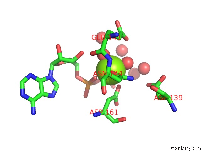
Mono view
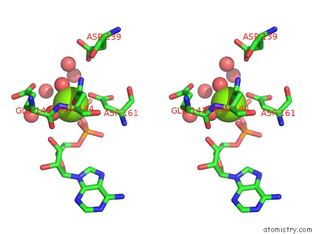
Stereo pair view
Mono view
Stereo pair view
A full contact list of Magnesium with other atoms in the Mg binding
site number 1 of X-Ray Structure of A Dap-Kinase 2-277 within 5.0Å range:
|
Reference:
I.De Diego,
J.Kuper,
F.Lehmann,
M.Wilmanns.
X-Ray Structure of A Dap-Kinase Calmodulin Complex To Be Published.
Page generated: Sun Aug 10 15:35:49 2025
Last articles
Mg in 6CA4Mg in 6C90
Mg in 6CA0
Mg in 6C9Y
Mg in 6C8Z
Mg in 6C8P
Mg in 6C8N
Mg in 6C8O
Mg in 6C8D
Mg in 6C8L