Magnesium »
PDB 2vu9-2wb1 »
2w5x »
Magnesium in PDB 2w5x: Structure of TAB5 Alkaline Phosphatase Mutant His 135 Glu with Mg Bound in the M3 Site.
Enzymatic activity of Structure of TAB5 Alkaline Phosphatase Mutant His 135 Glu with Mg Bound in the M3 Site.
All present enzymatic activity of Structure of TAB5 Alkaline Phosphatase Mutant His 135 Glu with Mg Bound in the M3 Site.:
3.1.3.1;
3.1.3.1;
Protein crystallography data
The structure of Structure of TAB5 Alkaline Phosphatase Mutant His 135 Glu with Mg Bound in the M3 Site., PDB code: 2w5x
was solved by
D.Koutsioulis,
A.Lyskowski,
S.Maki,
E.Guthrie,
G.Feller,
V.Bouriotis,
P.Heikinheimo,
with X-Ray Crystallography technique. A brief refinement statistics is given in the table below:
| Resolution Low / High (Å) | 44.65 / 1.99 |
| Space group | P 21 21 2 |
| Cell size a, b, c (Å), α, β, γ (°) | 70.420, 173.190, 55.440, 90.00, 90.00, 90.00 |
| R / Rfree (%) | 16.1 / 20.4 |
Other elements in 2w5x:
The structure of Structure of TAB5 Alkaline Phosphatase Mutant His 135 Glu with Mg Bound in the M3 Site. also contains other interesting chemical elements:
| Zinc | (Zn) | 4 atoms |
Magnesium Binding Sites:
The binding sites of Magnesium atom in the Structure of TAB5 Alkaline Phosphatase Mutant His 135 Glu with Mg Bound in the M3 Site.
(pdb code 2w5x). This binding sites where shown within
5.0 Angstroms radius around Magnesium atom.
In total 4 binding sites of Magnesium where determined in the Structure of TAB5 Alkaline Phosphatase Mutant His 135 Glu with Mg Bound in the M3 Site., PDB code: 2w5x:
Jump to Magnesium binding site number: 1; 2; 3; 4;
In total 4 binding sites of Magnesium where determined in the Structure of TAB5 Alkaline Phosphatase Mutant His 135 Glu with Mg Bound in the M3 Site., PDB code: 2w5x:
Jump to Magnesium binding site number: 1; 2; 3; 4;
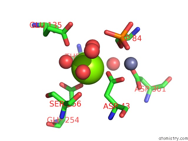
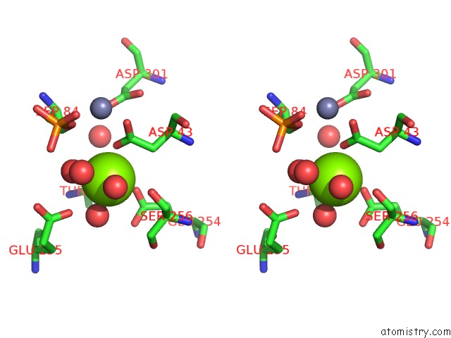
Magnesium binding site 1 out of 4 in 2w5x
Go back to
Magnesium binding site 1 out
of 4 in the Structure of TAB5 Alkaline Phosphatase Mutant His 135 Glu with Mg Bound in the M3 Site.
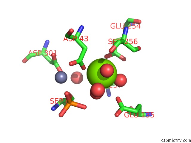
Mono view
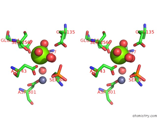
Stereo pair view
Mono view
Stereo pair view
A full contact list of Magnesium with other atoms in the Mg binding
site number 1 of Structure of TAB5 Alkaline Phosphatase Mutant His 135 Glu with Mg Bound in the M3 Site. within 5.0Å range:
|
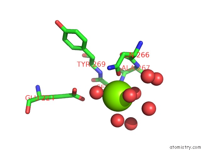
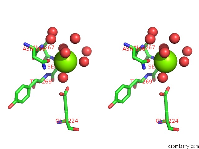
Magnesium binding site 2 out of 4 in 2w5x
Go back to
Magnesium binding site 2 out
of 4 in the Structure of TAB5 Alkaline Phosphatase Mutant His 135 Glu with Mg Bound in the M3 Site.
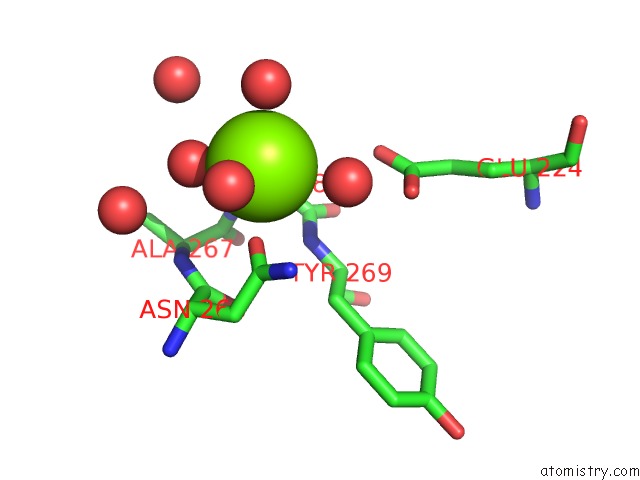
Mono view
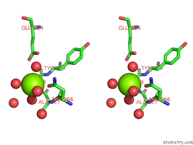
Stereo pair view
Mono view
Stereo pair view
A full contact list of Magnesium with other atoms in the Mg binding
site number 2 of Structure of TAB5 Alkaline Phosphatase Mutant His 135 Glu with Mg Bound in the M3 Site. within 5.0Å range:
|
Magnesium binding site 3 out of 4 in 2w5x
Go back to
Magnesium binding site 3 out
of 4 in the Structure of TAB5 Alkaline Phosphatase Mutant His 135 Glu with Mg Bound in the M3 Site.
Mono view
Stereo pair view
Mono view
Stereo pair view
A full contact list of Magnesium with other atoms in the Mg binding
site number 3 of Structure of TAB5 Alkaline Phosphatase Mutant His 135 Glu with Mg Bound in the M3 Site. within 5.0Å range:
|
Magnesium binding site 4 out of 4 in 2w5x
Go back to
Magnesium binding site 4 out
of 4 in the Structure of TAB5 Alkaline Phosphatase Mutant His 135 Glu with Mg Bound in the M3 Site.
Mono view
Stereo pair view
Mono view
Stereo pair view
A full contact list of Magnesium with other atoms in the Mg binding
site number 4 of Structure of TAB5 Alkaline Phosphatase Mutant His 135 Glu with Mg Bound in the M3 Site. within 5.0Å range:
|
Reference:
D.Koutsioulis,
A.Lyskowski,
S.Maki,
E.Guthrie,
G.Feller,
V.Bouriotis,
P.Heikinheimo.
Coordination Sphere of the Third Metal Site Is Essential to the Activity and Metal Selectivity of Alkaline Phosphatases. Protein Sci. V. 19 75 2010.
ISSN: ISSN 0961-8368
PubMed: 19916164
DOI: 10.1002/PRO.284
Page generated: Wed Aug 14 05:58:53 2024
ISSN: ISSN 0961-8368
PubMed: 19916164
DOI: 10.1002/PRO.284
Last articles
Zn in 9MJ5Zn in 9HNW
Zn in 9G0L
Zn in 9FNE
Zn in 9DZN
Zn in 9E0I
Zn in 9D32
Zn in 9DAK
Zn in 8ZXC
Zn in 8ZUF