Magnesium »
PDB 2wb4-2wkk »
2wfj »
Magnesium in PDB 2wfj: Atomic Resolution Crystal Structure of the Ppiase Domain of Human Cyclophilin G in Complex with Cyclosporin A.
Enzymatic activity of Atomic Resolution Crystal Structure of the Ppiase Domain of Human Cyclophilin G in Complex with Cyclosporin A.
All present enzymatic activity of Atomic Resolution Crystal Structure of the Ppiase Domain of Human Cyclophilin G in Complex with Cyclosporin A.:
5.2.1.8;
5.2.1.8;
Protein crystallography data
The structure of Atomic Resolution Crystal Structure of the Ppiase Domain of Human Cyclophilin G in Complex with Cyclosporin A., PDB code: 2wfj
was solved by
C.M.Stegmann,
G.M.Sheldrick,
M.C.Wahl,
with X-Ray Crystallography technique. A brief refinement statistics is given in the table below:
| Resolution Low / High (Å) | 10.00 / 0.75 |
| Space group | P 21 21 21 |
| Cell size a, b, c (Å), α, β, γ (°) | 37.319, 64.913, 69.285, 90.00, 90.00, 90.00 |
| R / Rfree (%) | 11.1 / 12.9 |
Other elements in 2wfj:
The structure of Atomic Resolution Crystal Structure of the Ppiase Domain of Human Cyclophilin G in Complex with Cyclosporin A. also contains other interesting chemical elements:
| Chlorine | (Cl) | 1 atom |
Magnesium Binding Sites:
The binding sites of Magnesium atom in the Atomic Resolution Crystal Structure of the Ppiase Domain of Human Cyclophilin G in Complex with Cyclosporin A.
(pdb code 2wfj). This binding sites where shown within
5.0 Angstroms radius around Magnesium atom.
In total 2 binding sites of Magnesium where determined in the Atomic Resolution Crystal Structure of the Ppiase Domain of Human Cyclophilin G in Complex with Cyclosporin A., PDB code: 2wfj:
Jump to Magnesium binding site number: 1; 2;
In total 2 binding sites of Magnesium where determined in the Atomic Resolution Crystal Structure of the Ppiase Domain of Human Cyclophilin G in Complex with Cyclosporin A., PDB code: 2wfj:
Jump to Magnesium binding site number: 1; 2;
Magnesium binding site 1 out of 2 in 2wfj
Go back to
Magnesium binding site 1 out
of 2 in the Atomic Resolution Crystal Structure of the Ppiase Domain of Human Cyclophilin G in Complex with Cyclosporin A.
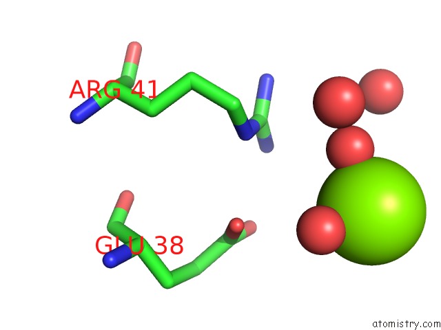
Mono view
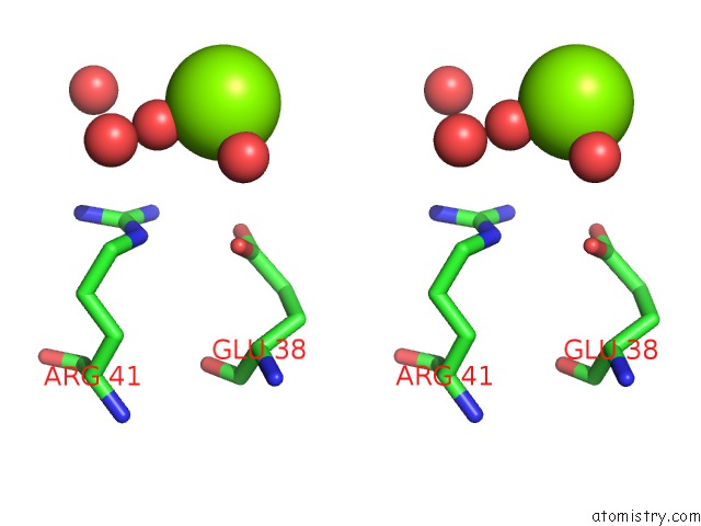
Stereo pair view
Mono view
Stereo pair view
A full contact list of Magnesium with other atoms in the Mg binding
site number 1 of Atomic Resolution Crystal Structure of the Ppiase Domain of Human Cyclophilin G in Complex with Cyclosporin A. within 5.0Å range:
|
Magnesium binding site 2 out of 2 in 2wfj
Go back to
Magnesium binding site 2 out
of 2 in the Atomic Resolution Crystal Structure of the Ppiase Domain of Human Cyclophilin G in Complex with Cyclosporin A.
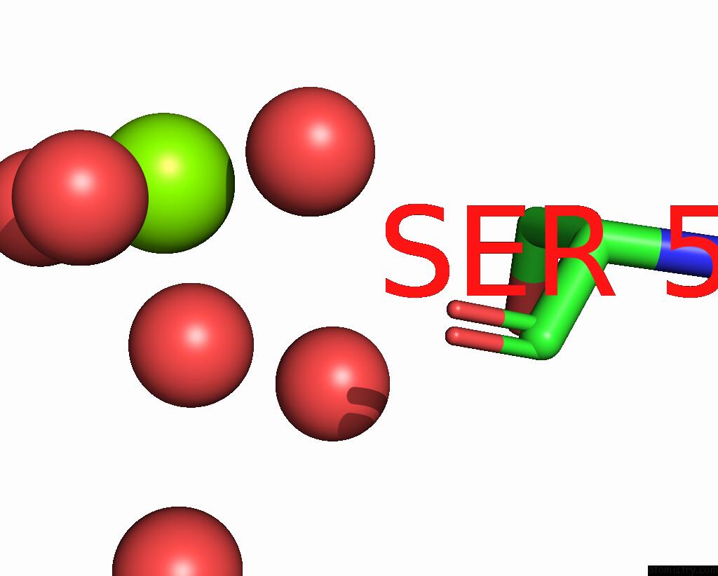
Mono view
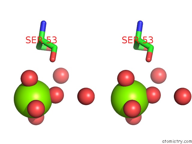
Stereo pair view
Mono view
Stereo pair view
A full contact list of Magnesium with other atoms in the Mg binding
site number 2 of Atomic Resolution Crystal Structure of the Ppiase Domain of Human Cyclophilin G in Complex with Cyclosporin A. within 5.0Å range:
|
Reference:
C.M.Stegmann,
D.Seeliger,
G.M.Sheldrick,
B.L.De Groot,
M.C.Wahl.
The Thermodynamic Influence of Trapped Water Molecules on A Protein-Ligand Interaction. Angew.Chem.Int.Ed.Engl. V. 48 5207 2009.
ISSN: ISSN 1433-7851
PubMed: 19499554
DOI: 10.1002/ANIE.200900481
Page generated: Sun Aug 10 15:41:33 2025
ISSN: ISSN 1433-7851
PubMed: 19499554
DOI: 10.1002/ANIE.200900481
Last articles
Mg in 6YIBMg in 6YIA
Mg in 6YBS
Mg in 6YHN
Mg in 6YHM
Mg in 6YF4
Mg in 6YEM
Mg in 6YE9
Mg in 6YDM
Mg in 6YDL