Magnesium »
PDB 2wb4-2wkk »
2wjh »
Magnesium in PDB 2wjh: Structure and Function of the Feob G-Domain From Methanococcus Jannaschii
Protein crystallography data
The structure of Structure and Function of the Feob G-Domain From Methanococcus Jannaschii, PDB code: 2wjh
was solved by
S.Koester,
M.Wehner,
C.Herrmann,
W.Kuehlbrandt,
O.Yildiz,
with X-Ray Crystallography technique. A brief refinement statistics is given in the table below:
| Resolution Low / High (Å) | 42.944 / 2.10 |
| Space group | C 1 2 1 |
| Cell size a, b, c (Å), α, β, γ (°) | 104.200, 50.400, 94.700, 90.00, 120.60, 90.00 |
| R / Rfree (%) | 21.04 / 24.61 |
Magnesium Binding Sites:
The binding sites of Magnesium atom in the Structure and Function of the Feob G-Domain From Methanococcus Jannaschii
(pdb code 2wjh). This binding sites where shown within
5.0 Angstroms radius around Magnesium atom.
In total 3 binding sites of Magnesium where determined in the Structure and Function of the Feob G-Domain From Methanococcus Jannaschii, PDB code: 2wjh:
Jump to Magnesium binding site number: 1; 2; 3;
In total 3 binding sites of Magnesium where determined in the Structure and Function of the Feob G-Domain From Methanococcus Jannaschii, PDB code: 2wjh:
Jump to Magnesium binding site number: 1; 2; 3;
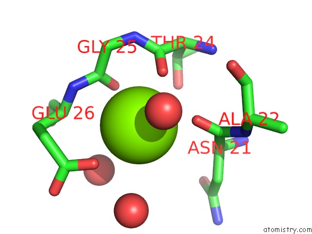
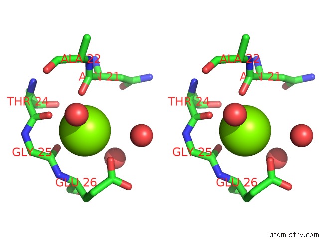
Magnesium binding site 1 out of 3 in 2wjh
Go back to
Magnesium binding site 1 out
of 3 in the Structure and Function of the Feob G-Domain From Methanococcus Jannaschii
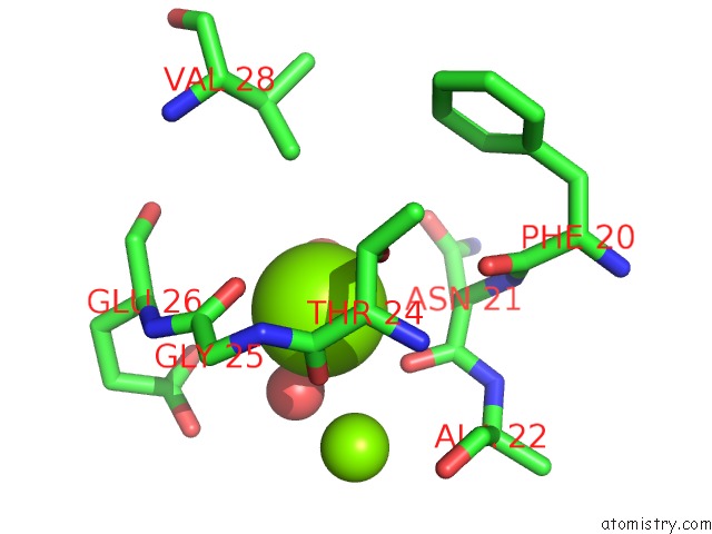
Mono view
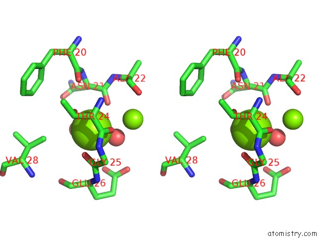
Stereo pair view
Mono view
Stereo pair view
A full contact list of Magnesium with other atoms in the Mg binding
site number 1 of Structure and Function of the Feob G-Domain From Methanococcus Jannaschii within 5.0Å range:
|
Magnesium binding site 2 out of 3 in 2wjh
Go back to
Magnesium binding site 2 out
of 3 in the Structure and Function of the Feob G-Domain From Methanococcus Jannaschii
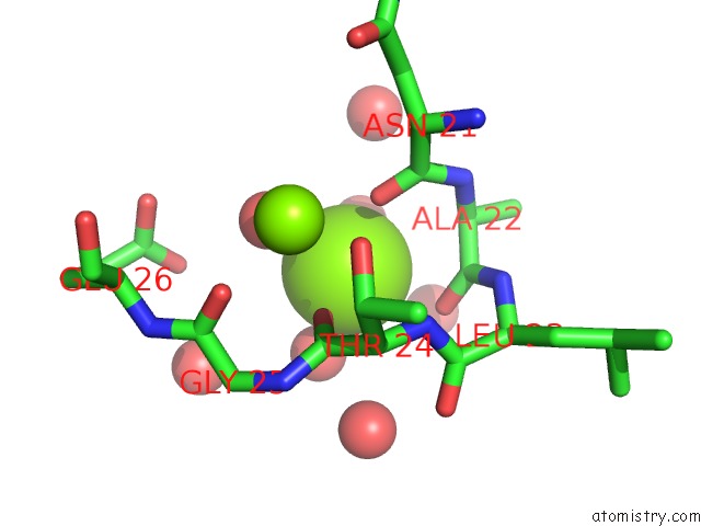
Mono view
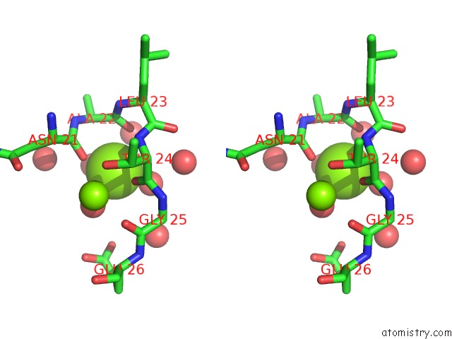
Stereo pair view
Mono view
Stereo pair view
A full contact list of Magnesium with other atoms in the Mg binding
site number 2 of Structure and Function of the Feob G-Domain From Methanococcus Jannaschii within 5.0Å range:
|
Magnesium binding site 3 out of 3 in 2wjh
Go back to
Magnesium binding site 3 out
of 3 in the Structure and Function of the Feob G-Domain From Methanococcus Jannaschii
Mono view
Stereo pair view
Mono view
Stereo pair view
A full contact list of Magnesium with other atoms in the Mg binding
site number 3 of Structure and Function of the Feob G-Domain From Methanococcus Jannaschii within 5.0Å range:
|
Reference:
S.Koester,
M.Wehner,
C.Herrmann,
W.Kuehlbrandt,
O.Yildiz.
Structure and Function of the Feob G-Domain From Methanococcus Jannaschii J.Mol.Biol. V. 392 405 2009.
ISSN: ISSN 0022-2836
PubMed: 19615379
DOI: 10.1016/J.JMB.2009.07.020
Page generated: Sun Aug 10 15:42:25 2025
ISSN: ISSN 0022-2836
PubMed: 19615379
DOI: 10.1016/J.JMB.2009.07.020
Last articles
Mg in 6YIAMg in 6YBS
Mg in 6YHN
Mg in 6YHM
Mg in 6YF4
Mg in 6YEM
Mg in 6YE9
Mg in 6YDM
Mg in 6YDL
Mg in 6YDJ