Magnesium »
PDB 2wkp-2x0q »
2ww8 »
Magnesium in PDB 2ww8: Structure of the Pilus Adhesin (Rrga) From Streptococcus Pneumoniae
Protein crystallography data
The structure of Structure of the Pilus Adhesin (Rrga) From Streptococcus Pneumoniae, PDB code: 2ww8
was solved by
T.Izore,
C.Contreras-Martel,
L.El-Mortaji,
C.Manzano,
R.Terrasse,
T.Vernet,
A.M.Di-Guilmi,
A.Dessen,
with X-Ray Crystallography technique. A brief refinement statistics is given in the table below:
| Resolution Low / High (Å) | 149.07 / 1.90 |
| Space group | P 21 21 21 |
| Cell size a, b, c (Å), α, β, γ (°) | 51.081, 82.700, 299.338, 90.00, 90.00, 90.00 |
| R / Rfree (%) | 19.518 / 23.46 |
Other elements in 2ww8:
The structure of Structure of the Pilus Adhesin (Rrga) From Streptococcus Pneumoniae also contains other interesting chemical elements:
| Calcium | (Ca) | 2 atoms |
Magnesium Binding Sites:
The binding sites of Magnesium atom in the Structure of the Pilus Adhesin (Rrga) From Streptococcus Pneumoniae
(pdb code 2ww8). This binding sites where shown within
5.0 Angstroms radius around Magnesium atom.
In total only one binding site of Magnesium was determined in the Structure of the Pilus Adhesin (Rrga) From Streptococcus Pneumoniae, PDB code: 2ww8:
In total only one binding site of Magnesium was determined in the Structure of the Pilus Adhesin (Rrga) From Streptococcus Pneumoniae, PDB code: 2ww8:
Magnesium binding site 1 out of 1 in 2ww8
Go back to
Magnesium binding site 1 out
of 1 in the Structure of the Pilus Adhesin (Rrga) From Streptococcus Pneumoniae
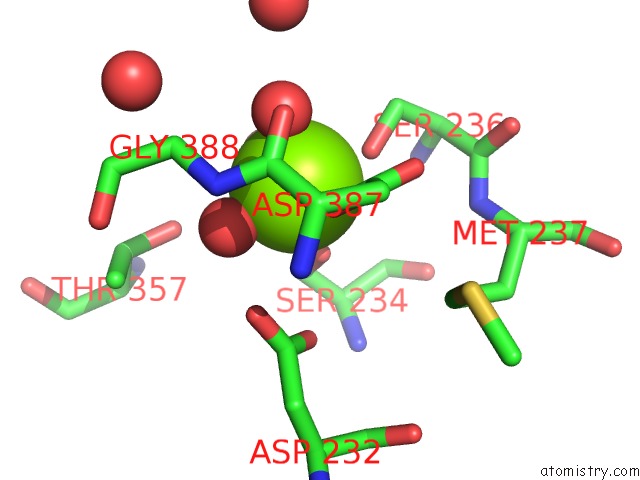
Mono view
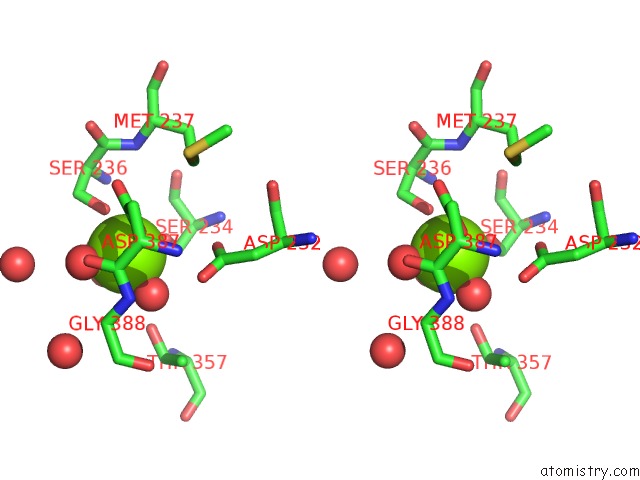
Stereo pair view
Mono view
Stereo pair view
A full contact list of Magnesium with other atoms in the Mg binding
site number 1 of Structure of the Pilus Adhesin (Rrga) From Streptococcus Pneumoniae within 5.0Å range:
|
Reference:
T.Izore,
C.Contreras-Martel,
L.El-Mortaji,
C.Manzano,
R.Terrasse,
T.Vernet,
A.M.Di-Guilmi,
A.Dessen.
Structural Basis of Host Cell Recognition By the Pilus Adhesin From Streptococcus Pneumoniae Structure V. 18 106 2010.
ISSN: ISSN 0969-2126
PubMed: 20152157
DOI: 10.1016/J.STR.2009.10.019
Page generated: Sun Aug 10 15:54:38 2025
ISSN: ISSN 0969-2126
PubMed: 20152157
DOI: 10.1016/J.STR.2009.10.019
Last articles
Mg in 4DPGMg in 4DQP
Mg in 4DQQ
Mg in 4DPM
Mg in 4DPV
Mg in 4DQI
Mg in 4DOB
Mg in 4DOC
Mg in 4DMZ
Mg in 4DOA