Magnesium »
PDB 2x13-2xbn »
2x5f »
Magnesium in PDB 2x5f: Crystal Structure of the Methicillin-Resistant Staphylococcus Aureus SAR2028, An ASPARTATE_TYROSINE_PHENYLALANINE Pyridoxal-5'-Phosphate Dependent Aminotransferase
Protein crystallography data
The structure of Crystal Structure of the Methicillin-Resistant Staphylococcus Aureus SAR2028, An ASPARTATE_TYROSINE_PHENYLALANINE Pyridoxal-5'-Phosphate Dependent Aminotransferase, PDB code: 2x5f
was solved by
M.Oke,
L.G.Carter,
K.A.Johnson,
H.Liu,
S.A.Mcmahon,
M.F.White,
J.H.Naismith,
with X-Ray Crystallography technique. A brief refinement statistics is given in the table below:
| Resolution Low / High (Å) | 27.24 / 1.80 |
| Space group | P 21 21 21 |
| Cell size a, b, c (Å), α, β, γ (°) | 82.780, 89.990, 104.910, 90.00, 90.00, 90.00 |
| R / Rfree (%) | 19.099 / 21.049 |
Magnesium Binding Sites:
The binding sites of Magnesium atom in the Crystal Structure of the Methicillin-Resistant Staphylococcus Aureus SAR2028, An ASPARTATE_TYROSINE_PHENYLALANINE Pyridoxal-5'-Phosphate Dependent Aminotransferase
(pdb code 2x5f). This binding sites where shown within
5.0 Angstroms radius around Magnesium atom.
In total only one binding site of Magnesium was determined in the Crystal Structure of the Methicillin-Resistant Staphylococcus Aureus SAR2028, An ASPARTATE_TYROSINE_PHENYLALANINE Pyridoxal-5'-Phosphate Dependent Aminotransferase, PDB code: 2x5f:
In total only one binding site of Magnesium was determined in the Crystal Structure of the Methicillin-Resistant Staphylococcus Aureus SAR2028, An ASPARTATE_TYROSINE_PHENYLALANINE Pyridoxal-5'-Phosphate Dependent Aminotransferase, PDB code: 2x5f:
Magnesium binding site 1 out of 1 in 2x5f
Go back to
Magnesium binding site 1 out
of 1 in the Crystal Structure of the Methicillin-Resistant Staphylococcus Aureus SAR2028, An ASPARTATE_TYROSINE_PHENYLALANINE Pyridoxal-5'-Phosphate Dependent Aminotransferase
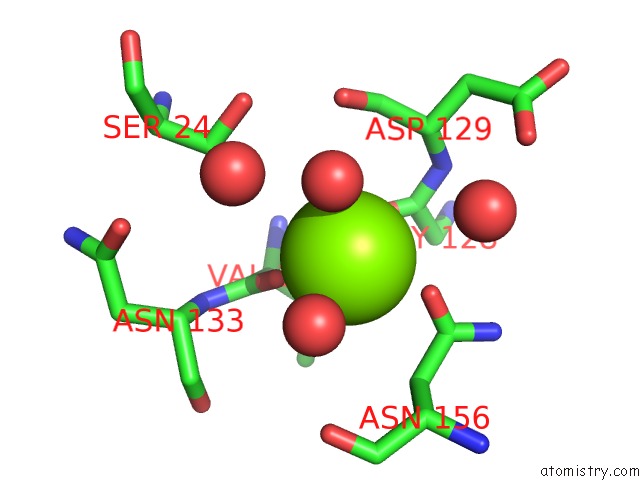
Mono view
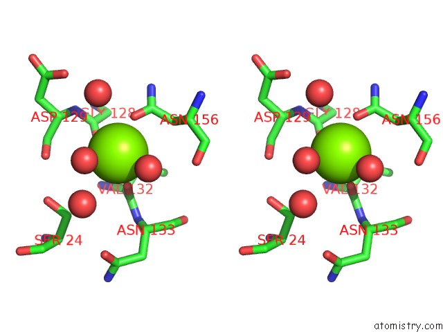
Stereo pair view
Mono view
Stereo pair view
A full contact list of Magnesium with other atoms in the Mg binding
site number 1 of Crystal Structure of the Methicillin-Resistant Staphylococcus Aureus SAR2028, An ASPARTATE_TYROSINE_PHENYLALANINE Pyridoxal-5'-Phosphate Dependent Aminotransferase within 5.0Å range:
|
Reference:
M.Oke,
L.G.Carter,
K.A.Johnson,
H.Liu,
S.A.Mcmahon,
X.Yan,
M.Kerou,
N.D.Weikart,
N.Kadi,
M.A.Sheikh,
S.Schmelz,
M.Dorward,
M.Zawadzki,
C.Cozens,
H.Falconer,
H.Powers,
I.M.Overton,
C.A.J.Van Niekerk,
X.Peng,
P.Patel,
R.A.Garrett,
D.Prangishvili,
C.H.Botting,
P.J.Coote,
D.T.F.Dryden,
G.J.Barton,
U.Schwarz-Linek,
G.L.Challis,
G.L.Taylor,
M.F.White,
J.H.Naismith.
The Scottish Structural Proteomics Facility: Targets, Methods and Outputs. J.Struct.Funct.Genom. V. 11 167 2010.
ISSN: ISSN 1345-711X
PubMed: 20419351
DOI: 10.1007/S10969-010-9090-Y
Page generated: Wed Aug 14 06:59:17 2024
ISSN: ISSN 1345-711X
PubMed: 20419351
DOI: 10.1007/S10969-010-9090-Y
Last articles
Fe in 2YXOFe in 2YRS
Fe in 2YXC
Fe in 2YNM
Fe in 2YVJ
Fe in 2YP1
Fe in 2YU2
Fe in 2YU1
Fe in 2YQB
Fe in 2YOO