Magnesium »
PDB 2x13-2xbn »
2x7c »
Magnesium in PDB 2x7c: Crystal Structure of Human Kinesin EG5 in Complex with (S)-Enastron
Protein crystallography data
The structure of Crystal Structure of Human Kinesin EG5 in Complex with (S)-Enastron, PDB code: 2x7c
was solved by
H.Y.K.Kaan,
V.Ulaganathan,
O.Rath,
C.Laggner,
H.Prokopcova,
D.Dallinger,
C.O.Kappe,
F.Kozielski,
with X-Ray Crystallography technique. A brief refinement statistics is given in the table below:
| Resolution Low / High (Å) | 30.00 / 1.90 |
| Space group | P 21 21 21 |
| Cell size a, b, c (Å), α, β, γ (°) | 69.620, 79.760, 159.330, 90.00, 90.00, 90.00 |
| R / Rfree (%) | 16.315 / 23.161 |
Magnesium Binding Sites:
The binding sites of Magnesium atom in the Crystal Structure of Human Kinesin EG5 in Complex with (S)-Enastron
(pdb code 2x7c). This binding sites where shown within
5.0 Angstroms radius around Magnesium atom.
In total 3 binding sites of Magnesium where determined in the Crystal Structure of Human Kinesin EG5 in Complex with (S)-Enastron, PDB code: 2x7c:
Jump to Magnesium binding site number: 1; 2; 3;
In total 3 binding sites of Magnesium where determined in the Crystal Structure of Human Kinesin EG5 in Complex with (S)-Enastron, PDB code: 2x7c:
Jump to Magnesium binding site number: 1; 2; 3;
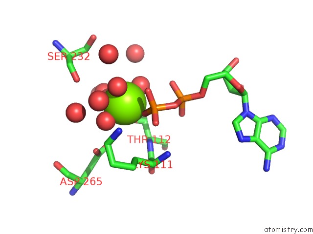
Magnesium binding site 1 out of 3 in 2x7c
Go back to
Magnesium binding site 1 out
of 3 in the Crystal Structure of Human Kinesin EG5 in Complex with (S)-Enastron
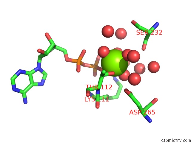
Mono view
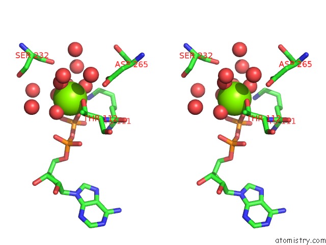
Stereo pair view
Mono view
Stereo pair view
A full contact list of Magnesium with other atoms in the Mg binding
site number 1 of Crystal Structure of Human Kinesin EG5 in Complex with (S)-Enastron within 5.0Å range:
|
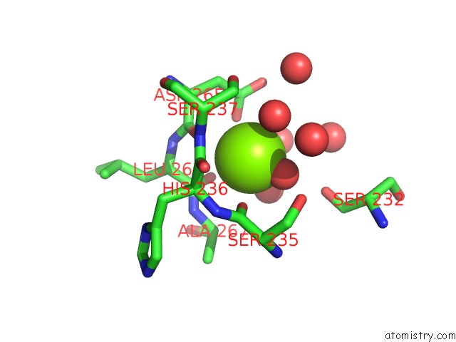
Magnesium binding site 2 out of 3 in 2x7c
Go back to
Magnesium binding site 2 out
of 3 in the Crystal Structure of Human Kinesin EG5 in Complex with (S)-Enastron
Mono view

Stereo pair view
Mono view
Stereo pair view
A full contact list of Magnesium with other atoms in the Mg binding
site number 2 of Crystal Structure of Human Kinesin EG5 in Complex with (S)-Enastron within 5.0Å range:
|
Magnesium binding site 3 out of 3 in 2x7c
Go back to
Magnesium binding site 3 out
of 3 in the Crystal Structure of Human Kinesin EG5 in Complex with (S)-Enastron
Mono view

Stereo pair view
Mono view
Stereo pair view
A full contact list of Magnesium with other atoms in the Mg binding
site number 3 of Crystal Structure of Human Kinesin EG5 in Complex with (S)-Enastron within 5.0Å range:
|
Reference:
H.Y.K.Kaan,
V.Ulaganathan,
O.Rath,
H.Prokopcova,
D.Dallinger,
C.O.Kappe,
F.Kozielski.
Structural Basis For Inhibition of EG5 By Dihydropyrimidines: Stereoselectivity of Antimitotic Inhibitors Enastron, Dimethylenastron and Fluorastrol. J.Med.Chem. V. 53 5676 2010.
ISSN: ISSN 0022-2623
PubMed: 20597485
DOI: 10.1021/JM100421N
Page generated: Wed Aug 14 07:01:18 2024
ISSN: ISSN 0022-2623
PubMed: 20597485
DOI: 10.1021/JM100421N
Last articles
Zn in 9J0NZn in 9J0O
Zn in 9J0P
Zn in 9FJX
Zn in 9EKB
Zn in 9C0F
Zn in 9CAH
Zn in 9CH0
Zn in 9CH3
Zn in 9CH1