Magnesium »
PDB 2x13-2xbn »
2xbj »
Magnesium in PDB 2xbj: Crystal Structure of CHK2 in Complex with An Inhibitor
Enzymatic activity of Crystal Structure of CHK2 in Complex with An Inhibitor
All present enzymatic activity of Crystal Structure of CHK2 in Complex with An Inhibitor:
2.7.11.1;
2.7.11.1;
Protein crystallography data
The structure of Crystal Structure of CHK2 in Complex with An Inhibitor, PDB code: 2xbj
was solved by
V.E.Anderson,
M.I.Walton,
P.D.Eve,
J.J.Caldwell,
L.H.Pearl,
A.W.Oliver,
I.Collins,
M.D.Garrett,
with X-Ray Crystallography technique. A brief refinement statistics is given in the table below:
| Resolution Low / High (Å) | 40.047 / 2.30 |
| Space group | P 32 2 1 |
| Cell size a, b, c (Å), α, β, γ (°) | 91.100, 91.100, 92.960, 90.00, 90.00, 120.00 |
| R / Rfree (%) | 19.37 / 24.33 |
Other elements in 2xbj:
The structure of Crystal Structure of CHK2 in Complex with An Inhibitor also contains other interesting chemical elements:
| Fluorine | (F) | 1 atom |
Magnesium Binding Sites:
The binding sites of Magnesium atom in the Crystal Structure of CHK2 in Complex with An Inhibitor
(pdb code 2xbj). This binding sites where shown within
5.0 Angstroms radius around Magnesium atom.
In total only one binding site of Magnesium was determined in the Crystal Structure of CHK2 in Complex with An Inhibitor, PDB code: 2xbj:
In total only one binding site of Magnesium was determined in the Crystal Structure of CHK2 in Complex with An Inhibitor, PDB code: 2xbj:
Magnesium binding site 1 out of 1 in 2xbj
Go back to
Magnesium binding site 1 out
of 1 in the Crystal Structure of CHK2 in Complex with An Inhibitor
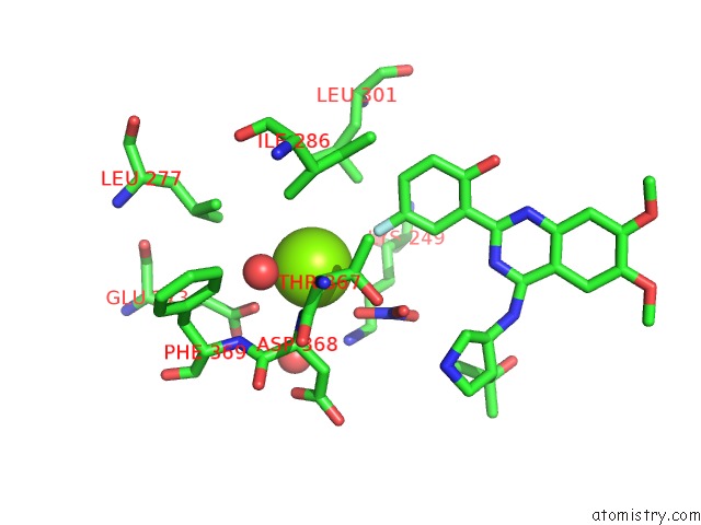
Mono view
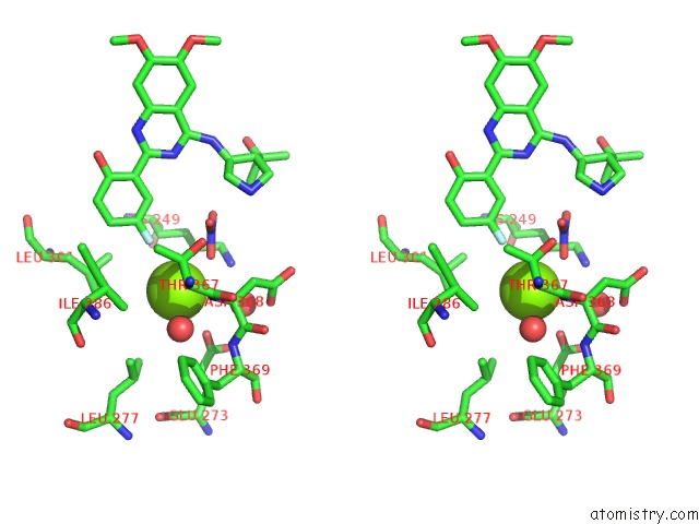
Stereo pair view
Mono view
Stereo pair view
A full contact list of Magnesium with other atoms in the Mg binding
site number 1 of Crystal Structure of CHK2 in Complex with An Inhibitor within 5.0Å range:
|
Reference:
J.J.Caldwell,
E.J.Welsh,
C.Matijssen,
V.E.Anderson,
L.Antoni,
K.Boxall,
F.Urban,
A.Hayes,
F.I.Raynaud,
L.J.Rigoreau,
T.Raynham,
G.W.Aherne,
L.H.Pearl,
A.W.Oliver,
M.D.Garrett,
I.Collins.
Structure-Based Design of Potent and Selective 2- (Quinazolin-2-Yl)Phenol Inhibitors of Checkpoint Kinase 2. J.Med.Chem. V. 54 580 2011.
ISSN: ISSN 0022-2623
PubMed: 21186793
DOI: 10.1021/JM101150B
Page generated: Wed Aug 14 07:03:50 2024
ISSN: ISSN 0022-2623
PubMed: 21186793
DOI: 10.1021/JM101150B
Last articles
Zn in 9J0NZn in 9J0O
Zn in 9J0P
Zn in 9FJX
Zn in 9EKB
Zn in 9C0F
Zn in 9CAH
Zn in 9CH0
Zn in 9CH3
Zn in 9CH1