Magnesium »
PDB 2xni-2xzs »
2xuu »
Magnesium in PDB 2xuu: Crystal Structure of A Dap-Kinase 1 Mutant
Enzymatic activity of Crystal Structure of A Dap-Kinase 1 Mutant
All present enzymatic activity of Crystal Structure of A Dap-Kinase 1 Mutant:
2.7.11.1;
2.7.11.1;
Protein crystallography data
The structure of Crystal Structure of A Dap-Kinase 1 Mutant, PDB code: 2xuu
was solved by
I.De Diego,
J.Kuper,
F.Lehmann,
M.Wilmanns,
with X-Ray Crystallography technique. A brief refinement statistics is given in the table below:
| Resolution Low / High (Å) | 60.36 / 1.80 |
| Space group | P 21 21 21 |
| Cell size a, b, c (Å), α, β, γ (°) | 49.740, 84.430, 86.340, 90.00, 90.00, 90.00 |
| R / Rfree (%) | 17.638 / 22.583 |
Magnesium Binding Sites:
The binding sites of Magnesium atom in the Crystal Structure of A Dap-Kinase 1 Mutant
(pdb code 2xuu). This binding sites where shown within
5.0 Angstroms radius around Magnesium atom.
In total only one binding site of Magnesium was determined in the Crystal Structure of A Dap-Kinase 1 Mutant, PDB code: 2xuu:
In total only one binding site of Magnesium was determined in the Crystal Structure of A Dap-Kinase 1 Mutant, PDB code: 2xuu:
Magnesium binding site 1 out of 1 in 2xuu
Go back to
Magnesium binding site 1 out
of 1 in the Crystal Structure of A Dap-Kinase 1 Mutant
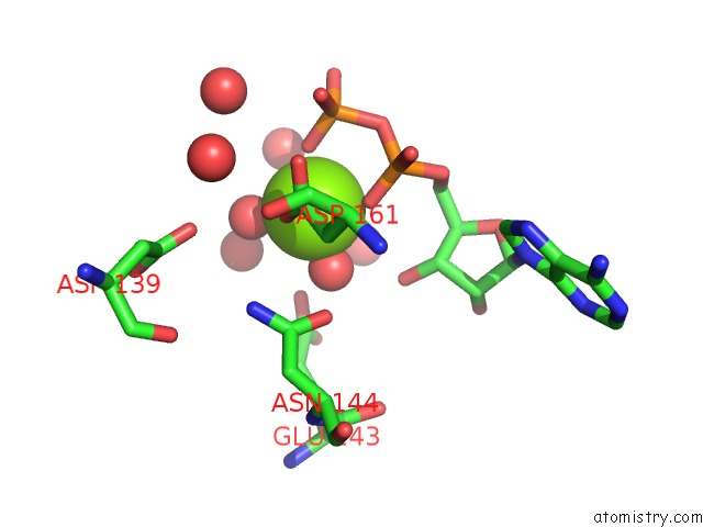
Mono view
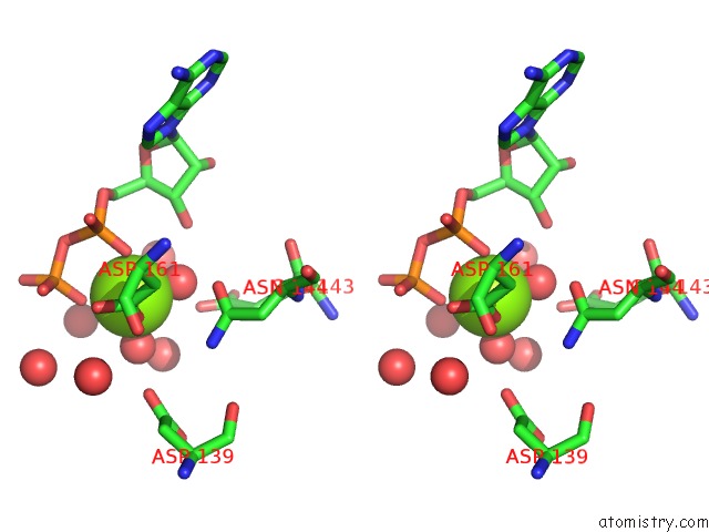
Stereo pair view
Mono view
Stereo pair view
A full contact list of Magnesium with other atoms in the Mg binding
site number 1 of Crystal Structure of A Dap-Kinase 1 Mutant within 5.0Å range:
|
Reference:
K.Temmerman,
I.De Diego,
V.Pogenberg,
B.Simon,
W.Jonko,
X.Li,
M.Wilmanns.
A Pef/Y Substrate Recognition and Signature Motif Plays A Critical Role in Dapk-Related Kinase Activity. Chem.Biol. V. 21 264 2014.
ISSN: ISSN 1074-5521
PubMed: 24440081
DOI: 10.1016/J.CHEMBIOL.2013.12.008
Page generated: Wed Aug 14 07:19:03 2024
ISSN: ISSN 1074-5521
PubMed: 24440081
DOI: 10.1016/J.CHEMBIOL.2013.12.008
Last articles
Zn in 9J0NZn in 9J0O
Zn in 9J0P
Zn in 9FJX
Zn in 9EKB
Zn in 9C0F
Zn in 9CAH
Zn in 9CH0
Zn in 9CH3
Zn in 9CH1