Magnesium »
PDB 2xni-2xzs »
2xy3 »
Magnesium in PDB 2xy3: Structure of the Bacillus Subtilis Prophage Dutpase with Dupnhpp
Enzymatic activity of Structure of the Bacillus Subtilis Prophage Dutpase with Dupnhpp
All present enzymatic activity of Structure of the Bacillus Subtilis Prophage Dutpase with Dupnhpp:
3.6.1.23;
3.6.1.23;
Protein crystallography data
The structure of Structure of the Bacillus Subtilis Prophage Dutpase with Dupnhpp, PDB code: 2xy3
was solved by
J.Garcia-Nafria,
M.Harkiolaki,
R.Persson,
M.J.Fogg,
K.S.Wilson,
with X-Ray Crystallography technique. A brief refinement statistics is given in the table below:
| Resolution Low / High (Å) | 20.00 / 2.55 |
| Space group | P 21 21 21 |
| Cell size a, b, c (Å), α, β, γ (°) | 98.474, 98.400, 97.993, 90.00, 90.00, 90.00 |
| R / Rfree (%) | 19.409 / 25.1 |
Magnesium Binding Sites:
The binding sites of Magnesium atom in the Structure of the Bacillus Subtilis Prophage Dutpase with Dupnhpp
(pdb code 2xy3). This binding sites where shown within
5.0 Angstroms radius around Magnesium atom.
In total 3 binding sites of Magnesium where determined in the Structure of the Bacillus Subtilis Prophage Dutpase with Dupnhpp, PDB code: 2xy3:
Jump to Magnesium binding site number: 1; 2; 3;
In total 3 binding sites of Magnesium where determined in the Structure of the Bacillus Subtilis Prophage Dutpase with Dupnhpp, PDB code: 2xy3:
Jump to Magnesium binding site number: 1; 2; 3;
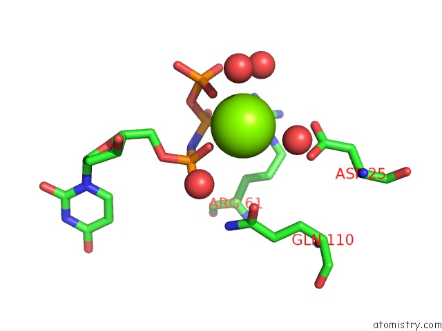
Magnesium binding site 1 out of 3 in 2xy3
Go back to
Magnesium binding site 1 out
of 3 in the Structure of the Bacillus Subtilis Prophage Dutpase with Dupnhpp
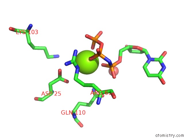
Mono view
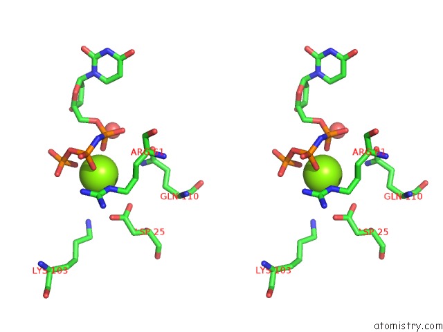
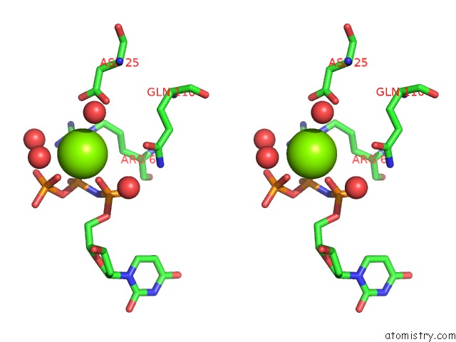
Stereo pair view
Mono view
Stereo pair view
A full contact list of Magnesium with other atoms in the Mg binding
site number 1 of Structure of the Bacillus Subtilis Prophage Dutpase with Dupnhpp within 5.0Å range:
|
Magnesium binding site 2 out of 3 in 2xy3
Go back to
Magnesium binding site 2 out
of 3 in the Structure of the Bacillus Subtilis Prophage Dutpase with Dupnhpp
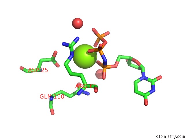
Mono view
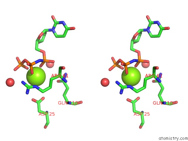
Stereo pair view
Mono view
Stereo pair view
A full contact list of Magnesium with other atoms in the Mg binding
site number 2 of Structure of the Bacillus Subtilis Prophage Dutpase with Dupnhpp within 5.0Å range:
|
Magnesium binding site 3 out of 3 in 2xy3
Go back to
Magnesium binding site 3 out
of 3 in the Structure of the Bacillus Subtilis Prophage Dutpase with Dupnhpp
Mono view
Stereo pair view
Mono view
Stereo pair view
A full contact list of Magnesium with other atoms in the Mg binding
site number 3 of Structure of the Bacillus Subtilis Prophage Dutpase with Dupnhpp within 5.0Å range:
|
Reference:
J.Garcia-Nafria,
M.Harkiolaki,
R.Persson,
M.J.Fogg,
K.S.Wilson.
The Structure of Bacillus Subtilis Sp Beta Prophage Dutpase and Its Complexes with Two Nucleotides Acta Crystallogr.,Sect.D V. 67 167 2011.
ISSN: ISSN 0907-4449
PubMed: 21358047
DOI: 10.1107/S0907444911003234
Page generated: Wed Aug 14 07:20:27 2024
ISSN: ISSN 0907-4449
PubMed: 21358047
DOI: 10.1107/S0907444911003234
Last articles
Zn in 9MJ5Zn in 9HNW
Zn in 9G0L
Zn in 9FNE
Zn in 9DZN
Zn in 9E0I
Zn in 9D32
Zn in 9DAK
Zn in 8ZXC
Zn in 8ZUF