Magnesium »
PDB 2ybb-2yni »
2ye1 »
Magnesium in PDB 2ye1: X-Ray Structure of the Cyan Fluorescent Proteinmturquoise-Gl (K206A Mutant)
Protein crystallography data
The structure of X-Ray Structure of the Cyan Fluorescent Proteinmturquoise-Gl (K206A Mutant), PDB code: 2ye1
was solved by
D.Von Stetten,
M.Noirclerc-Savoye,
J.Goedhart,
T.W.J.Gadella,
A.Royant,
with X-Ray Crystallography technique. A brief refinement statistics is given in the table below:
| Resolution Low / High (Å) | 41.26 / 1.63 |
| Space group | P 21 21 21 |
| Cell size a, b, c (Å), α, β, γ (°) | 51.140, 61.802, 69.814, 90.00, 90.00, 90.00 |
| R / Rfree (%) | 14.6 / 19.4 |
Magnesium Binding Sites:
The binding sites of Magnesium atom in the X-Ray Structure of the Cyan Fluorescent Proteinmturquoise-Gl (K206A Mutant)
(pdb code 2ye1). This binding sites where shown within
5.0 Angstroms radius around Magnesium atom.
In total only one binding site of Magnesium was determined in the X-Ray Structure of the Cyan Fluorescent Proteinmturquoise-Gl (K206A Mutant), PDB code: 2ye1:
In total only one binding site of Magnesium was determined in the X-Ray Structure of the Cyan Fluorescent Proteinmturquoise-Gl (K206A Mutant), PDB code: 2ye1:
Magnesium binding site 1 out of 1 in 2ye1
Go back to
Magnesium binding site 1 out
of 1 in the X-Ray Structure of the Cyan Fluorescent Proteinmturquoise-Gl (K206A Mutant)
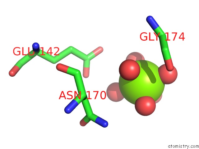
Mono view
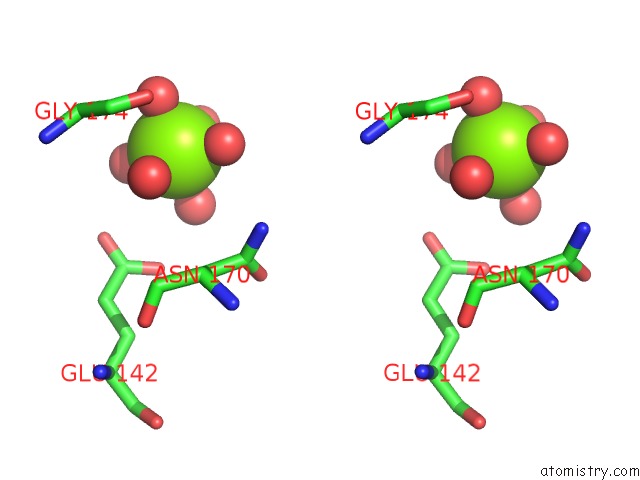
Stereo pair view
Mono view
Stereo pair view
A full contact list of Magnesium with other atoms in the Mg binding
site number 1 of X-Ray Structure of the Cyan Fluorescent Proteinmturquoise-Gl (K206A Mutant) within 5.0Å range:
|
Reference:
D.Von Stetten,
M.Noirclerc-Savoye,
J.Goedhart,
T.W.J.Gadella,
A.Royant.
Structural Characterization of the Cyan Fluorescent Protein Mturquoise-Gl To Be Published.
Page generated: Wed Aug 14 07:29:56 2024
Last articles
Zn in 9JYWZn in 9IR4
Zn in 9IR3
Zn in 9GMX
Zn in 9GMW
Zn in 9JEJ
Zn in 9ERF
Zn in 9ERE
Zn in 9EGV
Zn in 9EGW