Magnesium »
PDB 2ybb-2yni »
2yg1 »
Magnesium in PDB 2yg1: Apo Structure of Cellobiohydrolase 1 (CEL7A) From Heterobasidion Annosum
Protein crystallography data
The structure of Apo Structure of Cellobiohydrolase 1 (CEL7A) From Heterobasidion Annosum, PDB code: 2yg1
was solved by
M.Haddad-Momeni,
H.Hansson,
N.E.Mikkelsen,
X.Wang,
J.Svedberg,
M.Sandgren,
J.Stahlberg,
with X-Ray Crystallography technique. A brief refinement statistics is given in the table below:
| Resolution Low / High (Å) | 28.10 / 1.90 |
| Space group | P 1 21 1 |
| Cell size a, b, c (Å), α, β, γ (°) | 60.511, 84.103, 75.877, 90.00, 103.36, 90.00 |
| R / Rfree (%) | 19.7 / 25.3 |
Magnesium Binding Sites:
The binding sites of Magnesium atom in the Apo Structure of Cellobiohydrolase 1 (CEL7A) From Heterobasidion Annosum
(pdb code 2yg1). This binding sites where shown within
5.0 Angstroms radius around Magnesium atom.
In total 3 binding sites of Magnesium where determined in the Apo Structure of Cellobiohydrolase 1 (CEL7A) From Heterobasidion Annosum, PDB code: 2yg1:
Jump to Magnesium binding site number: 1; 2; 3;
In total 3 binding sites of Magnesium where determined in the Apo Structure of Cellobiohydrolase 1 (CEL7A) From Heterobasidion Annosum, PDB code: 2yg1:
Jump to Magnesium binding site number: 1; 2; 3;
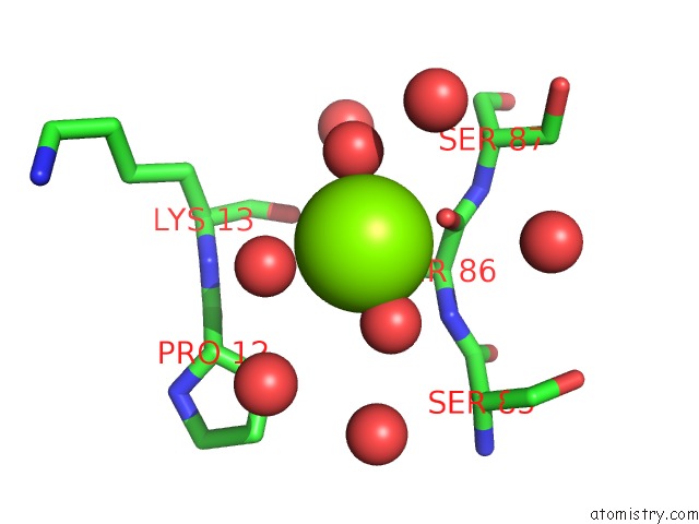
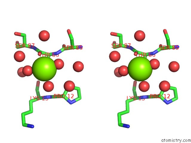
Magnesium binding site 1 out of 3 in 2yg1
Go back to
Magnesium binding site 1 out
of 3 in the Apo Structure of Cellobiohydrolase 1 (CEL7A) From Heterobasidion Annosum
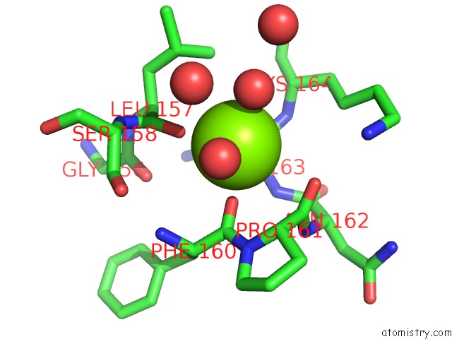
Mono view
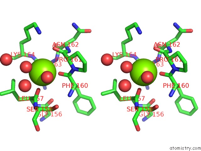
Stereo pair view
Mono view
Stereo pair view
A full contact list of Magnesium with other atoms in the Mg binding
site number 1 of Apo Structure of Cellobiohydrolase 1 (CEL7A) From Heterobasidion Annosum within 5.0Å range:
|
Magnesium binding site 2 out of 3 in 2yg1
Go back to
Magnesium binding site 2 out
of 3 in the Apo Structure of Cellobiohydrolase 1 (CEL7A) From Heterobasidion Annosum
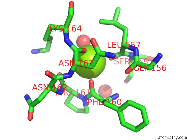
Mono view
Stereo pair view
Mono view
Stereo pair view
A full contact list of Magnesium with other atoms in the Mg binding
site number 2 of Apo Structure of Cellobiohydrolase 1 (CEL7A) From Heterobasidion Annosum within 5.0Å range:
|
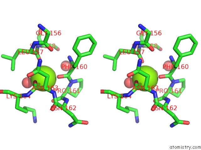
Magnesium binding site 3 out of 3 in 2yg1
Go back to
Magnesium binding site 3 out
of 3 in the Apo Structure of Cellobiohydrolase 1 (CEL7A) From Heterobasidion Annosum
Mono view
Stereo pair view
Mono view
Stereo pair view
A full contact list of Magnesium with other atoms in the Mg binding
site number 3 of Apo Structure of Cellobiohydrolase 1 (CEL7A) From Heterobasidion Annosum within 5.0Å range:
|
Reference:
M.H.Momeni,
C.M.Payne,
H.Hansson,
N.E.Mikkelsen,
J.Svedberg,
A.Engstrom,
M.Sandgren,
G.T.Beckham,
J.Stahlberg.
Structural, Biochemical, and Computational Characterization of the Glycoside Hydrolase Family 7 Cellobiohydrolase of the Tree-Killing Fungus Heterobasidion Irregulare. J.Biol.Chem. V. 288 5861 2013.
ISSN: ISSN 0021-9258
PubMed: 23303184
DOI: 10.1074/JBC.M112.440891
Page generated: Wed Aug 14 07:30:39 2024
ISSN: ISSN 0021-9258
PubMed: 23303184
DOI: 10.1074/JBC.M112.440891
Last articles
Zn in 9J0NZn in 9J0O
Zn in 9J0P
Zn in 9FJX
Zn in 9EKB
Zn in 9C0F
Zn in 9CAH
Zn in 9CH0
Zn in 9CH3
Zn in 9CH1