Magnesium »
PDB 2ynj-2z4w »
2z49 »
Magnesium in PDB 2z49: Crystal Structure of Hemolytic Lectin Cel-III Complexed with Methyl- Alpha-D-Galactopylanoside
Protein crystallography data
The structure of Crystal Structure of Hemolytic Lectin Cel-III Complexed with Methyl- Alpha-D-Galactopylanoside, PDB code: 2z49
was solved by
T.Hatakeyama,
H.Unno,
S.Eto,
H.Hidemura,
T.Uchida,
Y.Kouzuma,
with X-Ray Crystallography technique. A brief refinement statistics is given in the table below:
| Resolution Low / High (Å) | 58.12 / 1.95 |
| Space group | P 1 21 1 |
| Cell size a, b, c (Å), α, β, γ (°) | 53.287, 65.451, 126.993, 90.00, 97.07, 90.00 |
| R / Rfree (%) | 19.1 / 23.9 |
Other elements in 2z49:
The structure of Crystal Structure of Hemolytic Lectin Cel-III Complexed with Methyl- Alpha-D-Galactopylanoside also contains other interesting chemical elements:
| Calcium | (Ca) | 10 atoms |
Magnesium Binding Sites:
The binding sites of Magnesium atom in the Crystal Structure of Hemolytic Lectin Cel-III Complexed with Methyl- Alpha-D-Galactopylanoside
(pdb code 2z49). This binding sites where shown within
5.0 Angstroms radius around Magnesium atom.
In total 4 binding sites of Magnesium where determined in the Crystal Structure of Hemolytic Lectin Cel-III Complexed with Methyl- Alpha-D-Galactopylanoside, PDB code: 2z49:
Jump to Magnesium binding site number: 1; 2; 3; 4;
In total 4 binding sites of Magnesium where determined in the Crystal Structure of Hemolytic Lectin Cel-III Complexed with Methyl- Alpha-D-Galactopylanoside, PDB code: 2z49:
Jump to Magnesium binding site number: 1; 2; 3; 4;
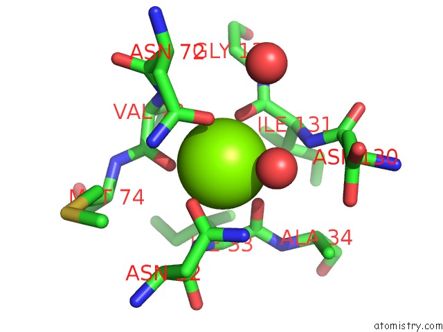
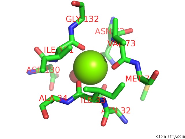
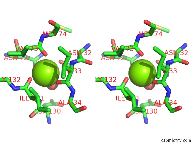
Magnesium binding site 1 out of 4 in 2z49
Go back to
Magnesium binding site 1 out
of 4 in the Crystal Structure of Hemolytic Lectin Cel-III Complexed with Methyl- Alpha-D-Galactopylanoside
Mono view
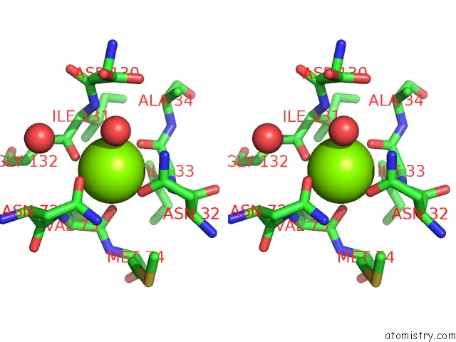
Stereo pair view
Mono view
Stereo pair view
A full contact list of Magnesium with other atoms in the Mg binding
site number 1 of Crystal Structure of Hemolytic Lectin Cel-III Complexed with Methyl- Alpha-D-Galactopylanoside within 5.0Å range:
|
Magnesium binding site 2 out of 4 in 2z49
Go back to
Magnesium binding site 2 out
of 4 in the Crystal Structure of Hemolytic Lectin Cel-III Complexed with Methyl- Alpha-D-Galactopylanoside
Mono view
Stereo pair view
Mono view
Stereo pair view
A full contact list of Magnesium with other atoms in the Mg binding
site number 2 of Crystal Structure of Hemolytic Lectin Cel-III Complexed with Methyl- Alpha-D-Galactopylanoside within 5.0Å range:
|
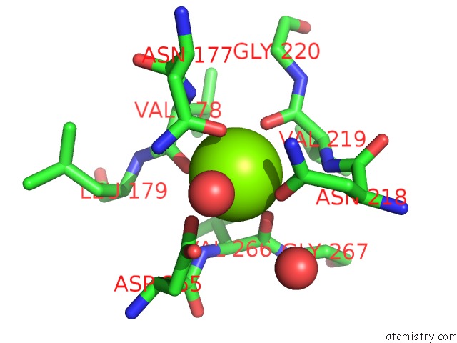
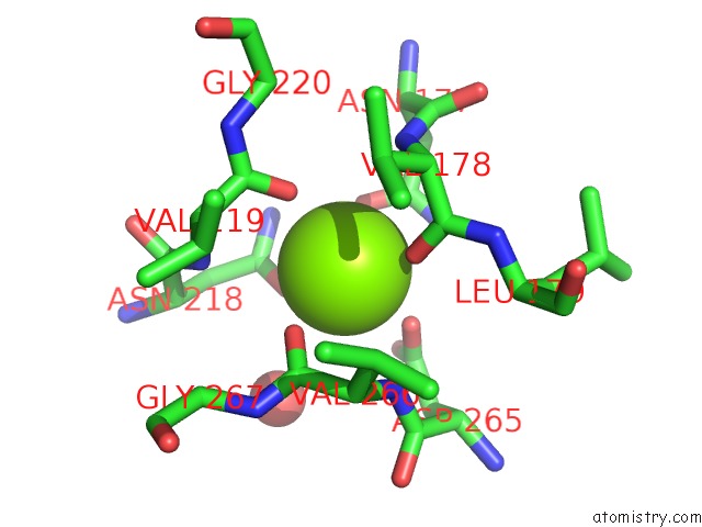
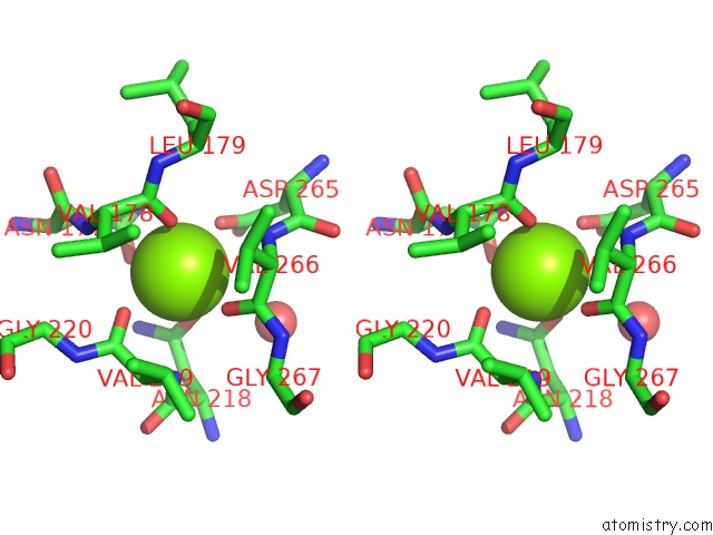
Magnesium binding site 3 out of 4 in 2z49
Go back to
Magnesium binding site 3 out
of 4 in the Crystal Structure of Hemolytic Lectin Cel-III Complexed with Methyl- Alpha-D-Galactopylanoside
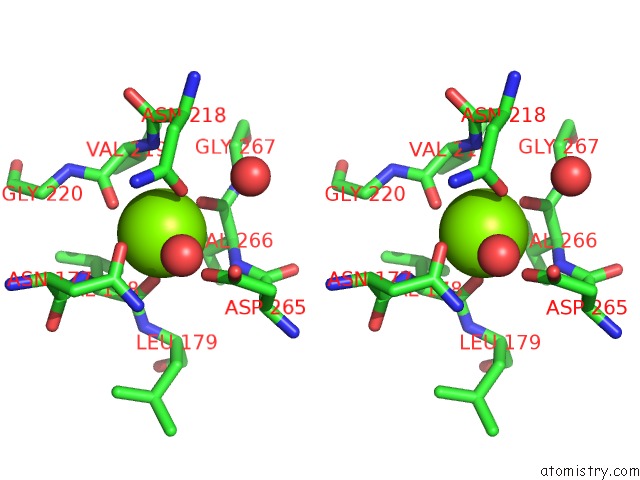
Mono view
Stereo pair view
Mono view
Stereo pair view
A full contact list of Magnesium with other atoms in the Mg binding
site number 3 of Crystal Structure of Hemolytic Lectin Cel-III Complexed with Methyl- Alpha-D-Galactopylanoside within 5.0Å range:
|
Magnesium binding site 4 out of 4 in 2z49
Go back to
Magnesium binding site 4 out
of 4 in the Crystal Structure of Hemolytic Lectin Cel-III Complexed with Methyl- Alpha-D-Galactopylanoside
Mono view
Stereo pair view
Mono view
Stereo pair view
A full contact list of Magnesium with other atoms in the Mg binding
site number 4 of Crystal Structure of Hemolytic Lectin Cel-III Complexed with Methyl- Alpha-D-Galactopylanoside within 5.0Å range:
|
Reference:
T.Hatakeyama,
H.Unno,
Y.Kouzuma,
T.Uchida,
S.Eto,
H.Hidemura,
N.Kato,
M.Yonekura,
M.Kusunoki.
C-Type Lectin-Like Carbohydrate-Recognition of the Hemolytic Lectin Cel-III Containing Ricin-Type Beta-Trefoil Folds J.Biol.Chem. V. 282 37826 2007.
ISSN: ISSN 0021-9258
PubMed: 17977832
DOI: 10.1074/JBC.M705604200
Page generated: Wed Aug 14 07:44:03 2024
ISSN: ISSN 0021-9258
PubMed: 17977832
DOI: 10.1074/JBC.M705604200
Last articles
Fe in 2YXOFe in 2YRS
Fe in 2YXC
Fe in 2YNM
Fe in 2YVJ
Fe in 2YP1
Fe in 2YU2
Fe in 2YU1
Fe in 2YQB
Fe in 2YOO