Magnesium »
PDB 2z4x-2zin »
2z7q »
Magnesium in PDB 2z7q: Crystal Structure of the N-Terminal Kinase Domain of Human Rsk-1 Bound to Amp-Pcp
Enzymatic activity of Crystal Structure of the N-Terminal Kinase Domain of Human Rsk-1 Bound to Amp-Pcp
All present enzymatic activity of Crystal Structure of the N-Terminal Kinase Domain of Human Rsk-1 Bound to Amp-Pcp:
2.7.11.1;
2.7.11.1;
Protein crystallography data
The structure of Crystal Structure of the N-Terminal Kinase Domain of Human Rsk-1 Bound to Amp-Pcp, PDB code: 2z7q
was solved by
M.Ikuta,
S.K.Munshi,
with X-Ray Crystallography technique. A brief refinement statistics is given in the table below:
| Resolution Low / High (Å) | 20.00 / 2.00 |
| Space group | P 21 21 21 |
| Cell size a, b, c (Å), α, β, γ (°) | 52.194, 53.685, 119.874, 90.00, 90.00, 90.00 |
| R / Rfree (%) | n/a / n/a |
Magnesium Binding Sites:
The binding sites of Magnesium atom in the Crystal Structure of the N-Terminal Kinase Domain of Human Rsk-1 Bound to Amp-Pcp
(pdb code 2z7q). This binding sites where shown within
5.0 Angstroms radius around Magnesium atom.
In total only one binding site of Magnesium was determined in the Crystal Structure of the N-Terminal Kinase Domain of Human Rsk-1 Bound to Amp-Pcp, PDB code: 2z7q:
In total only one binding site of Magnesium was determined in the Crystal Structure of the N-Terminal Kinase Domain of Human Rsk-1 Bound to Amp-Pcp, PDB code: 2z7q:
Magnesium binding site 1 out of 1 in 2z7q
Go back to
Magnesium binding site 1 out
of 1 in the Crystal Structure of the N-Terminal Kinase Domain of Human Rsk-1 Bound to Amp-Pcp
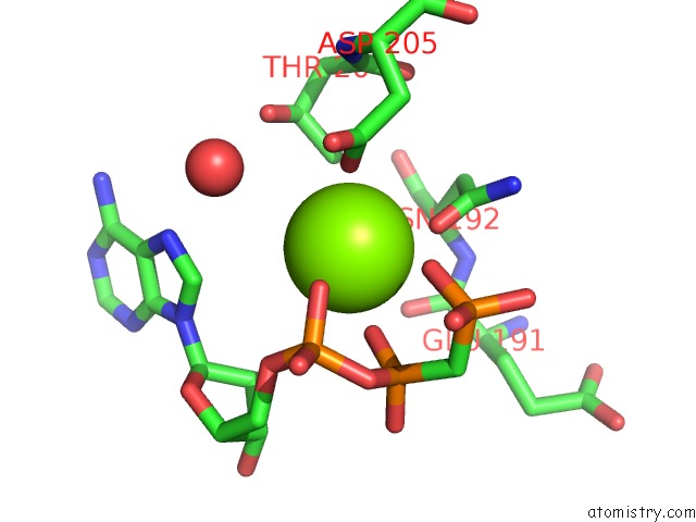
Mono view
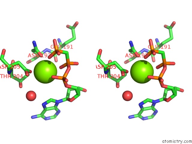
Stereo pair view
Mono view
Stereo pair view
A full contact list of Magnesium with other atoms in the Mg binding
site number 1 of Crystal Structure of the N-Terminal Kinase Domain of Human Rsk-1 Bound to Amp-Pcp within 5.0Å range:
|
Reference:
M.Ikuta,
M.Kornienko,
N.Byrne,
J.C.Reid,
S.Mizuarai,
H.Kotani,
S.K.Munshi.
Crystal Structures of the N-Terminal Kinase Domain of Human RSK1 Bound to Three Different Ligands: Implications For the Design of RSK1 Specific Inhibitors. Protein Sci. V. 16 2626 2007.
ISSN: ISSN 0961-8368
PubMed: 17965187
DOI: 10.1110/PS.073123707
Page generated: Wed Aug 14 07:48:02 2024
ISSN: ISSN 0961-8368
PubMed: 17965187
DOI: 10.1110/PS.073123707
Last articles
Zn in 9J0NZn in 9J0O
Zn in 9J0P
Zn in 9FJX
Zn in 9EKB
Zn in 9C0F
Zn in 9CAH
Zn in 9CH0
Zn in 9CH3
Zn in 9CH1