Magnesium »
PDB 2zjp-2zvj »
2zut »
Magnesium in PDB 2zut: Crystal Structure of Galacto-N-Biose/Lacto-N-Biose I Phosphorylase in Complex with Galnac
Enzymatic activity of Crystal Structure of Galacto-N-Biose/Lacto-N-Biose I Phosphorylase in Complex with Galnac
All present enzymatic activity of Crystal Structure of Galacto-N-Biose/Lacto-N-Biose I Phosphorylase in Complex with Galnac:
2.4.1.211;
2.4.1.211;
Protein crystallography data
The structure of Crystal Structure of Galacto-N-Biose/Lacto-N-Biose I Phosphorylase in Complex with Galnac, PDB code: 2zut
was solved by
M.Hidaka,
M.Nishimoto,
M.Kitaoka,
T.Wakagi,
H.Shoun,
S.Fushinobu,
with X-Ray Crystallography technique. A brief refinement statistics is given in the table below:
| Resolution Low / High (Å) | 44.81 / 1.90 |
| Space group | P 1 |
| Cell size a, b, c (Å), α, β, γ (°) | 67.861, 111.658, 118.660, 105.20, 90.48, 107.27 |
| R / Rfree (%) | 16.1 / 20.4 |
Magnesium Binding Sites:
The binding sites of Magnesium atom in the Crystal Structure of Galacto-N-Biose/Lacto-N-Biose I Phosphorylase in Complex with Galnac
(pdb code 2zut). This binding sites where shown within
5.0 Angstroms radius around Magnesium atom.
In total 6 binding sites of Magnesium where determined in the Crystal Structure of Galacto-N-Biose/Lacto-N-Biose I Phosphorylase in Complex with Galnac, PDB code: 2zut:
Jump to Magnesium binding site number: 1; 2; 3; 4; 5; 6;
In total 6 binding sites of Magnesium where determined in the Crystal Structure of Galacto-N-Biose/Lacto-N-Biose I Phosphorylase in Complex with Galnac, PDB code: 2zut:
Jump to Magnesium binding site number: 1; 2; 3; 4; 5; 6;
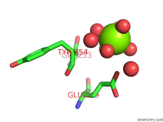
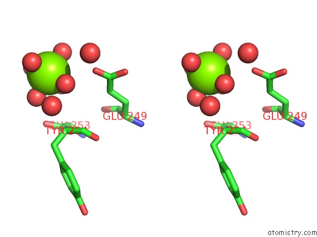
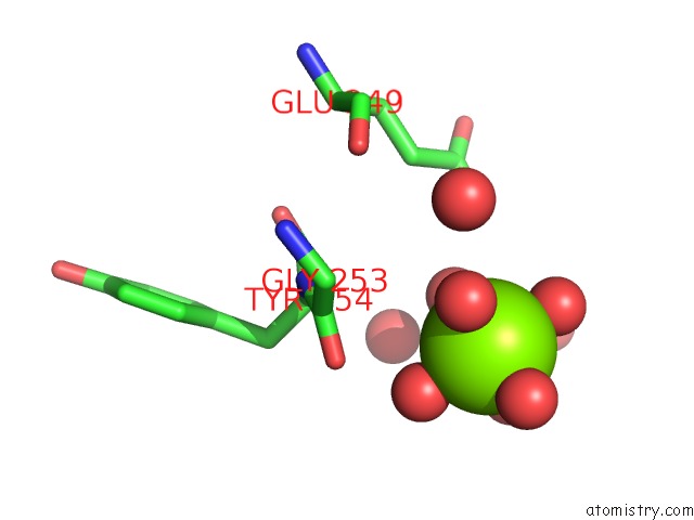
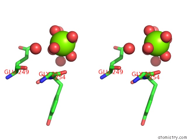
Magnesium binding site 1 out of 6 in 2zut
Go back to
Magnesium binding site 1 out
of 6 in the Crystal Structure of Galacto-N-Biose/Lacto-N-Biose I Phosphorylase in Complex with Galnac
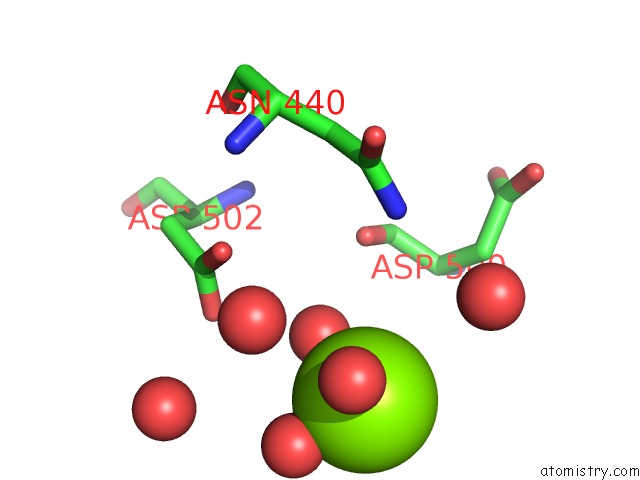
Mono view
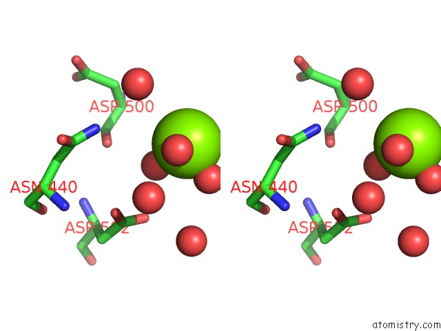
Stereo pair view
Mono view
Stereo pair view
A full contact list of Magnesium with other atoms in the Mg binding
site number 1 of Crystal Structure of Galacto-N-Biose/Lacto-N-Biose I Phosphorylase in Complex with Galnac within 5.0Å range:
|
Magnesium binding site 2 out of 6 in 2zut
Go back to
Magnesium binding site 2 out
of 6 in the Crystal Structure of Galacto-N-Biose/Lacto-N-Biose I Phosphorylase in Complex with Galnac
Mono view
Stereo pair view
Mono view
Stereo pair view
A full contact list of Magnesium with other atoms in the Mg binding
site number 2 of Crystal Structure of Galacto-N-Biose/Lacto-N-Biose I Phosphorylase in Complex with Galnac within 5.0Å range:
|
Magnesium binding site 3 out of 6 in 2zut
Go back to
Magnesium binding site 3 out
of 6 in the Crystal Structure of Galacto-N-Biose/Lacto-N-Biose I Phosphorylase in Complex with Galnac
Mono view
Stereo pair view
Mono view
Stereo pair view
A full contact list of Magnesium with other atoms in the Mg binding
site number 3 of Crystal Structure of Galacto-N-Biose/Lacto-N-Biose I Phosphorylase in Complex with Galnac within 5.0Å range:
|
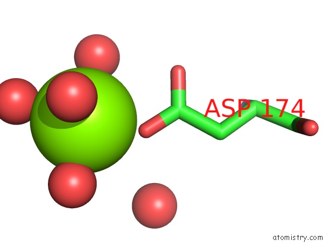
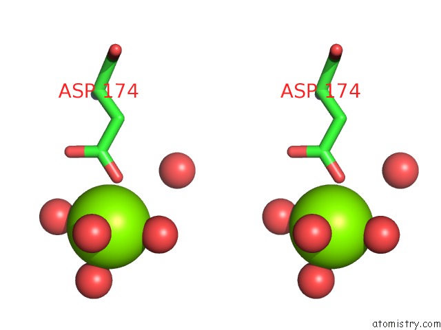
Magnesium binding site 4 out of 6 in 2zut
Go back to
Magnesium binding site 4 out
of 6 in the Crystal Structure of Galacto-N-Biose/Lacto-N-Biose I Phosphorylase in Complex with Galnac
Mono view
Stereo pair view
Mono view
Stereo pair view
A full contact list of Magnesium with other atoms in the Mg binding
site number 4 of Crystal Structure of Galacto-N-Biose/Lacto-N-Biose I Phosphorylase in Complex with Galnac within 5.0Å range:
|
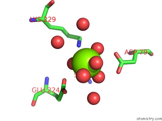
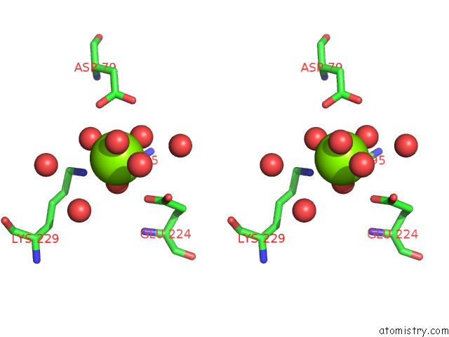
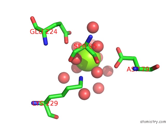
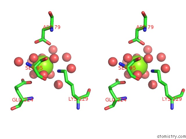
Magnesium binding site 5 out of 6 in 2zut
Go back to
Magnesium binding site 5 out
of 6 in the Crystal Structure of Galacto-N-Biose/Lacto-N-Biose I Phosphorylase in Complex with Galnac
Mono view
Stereo pair view
Mono view
Stereo pair view
A full contact list of Magnesium with other atoms in the Mg binding
site number 5 of Crystal Structure of Galacto-N-Biose/Lacto-N-Biose I Phosphorylase in Complex with Galnac within 5.0Å range:
|
Magnesium binding site 6 out of 6 in 2zut
Go back to
Magnesium binding site 6 out
of 6 in the Crystal Structure of Galacto-N-Biose/Lacto-N-Biose I Phosphorylase in Complex with Galnac
Mono view
Stereo pair view
Mono view
Stereo pair view
A full contact list of Magnesium with other atoms in the Mg binding
site number 6 of Crystal Structure of Galacto-N-Biose/Lacto-N-Biose I Phosphorylase in Complex with Galnac within 5.0Å range:
|
Reference:
M.Hidaka,
M.Nishimoto,
M.Kitaoka,
T.Wakagi,
H.Shoun,
S.Fushinobu.
The Crystal Structure of Galacto-N-Biose/Lacto-N-Biose I Phosphorylase: A Large Deformation of A Tim Barrel Scaffold J.Biol.Chem. V. 284 7273 2009.
ISSN: ISSN 0021-9258
PubMed: 19124470
DOI: 10.1074/JBC.M808525200
Page generated: Sun Aug 10 17:05:48 2025
ISSN: ISSN 0021-9258
PubMed: 19124470
DOI: 10.1074/JBC.M808525200
Last articles
Mg in 6C62Mg in 6C5U
Mg in 6C5N
Mg in 6C55
Mg in 6C2W
Mg in 6C3P
Mg in 6C3O
Mg in 6C2C
Mg in 6C2X
Mg in 6C25