Magnesium »
PDB 2zjp-2zvj »
2zvj »
Magnesium in PDB 2zvj: Crystal Structures of Rat Catechol-O-Methyltransferase Complexed with Coumarine-Based Inhibitor
Enzymatic activity of Crystal Structures of Rat Catechol-O-Methyltransferase Complexed with Coumarine-Based Inhibitor
All present enzymatic activity of Crystal Structures of Rat Catechol-O-Methyltransferase Complexed with Coumarine-Based Inhibitor:
2.1.1.6;
2.1.1.6;
Protein crystallography data
The structure of Crystal Structures of Rat Catechol-O-Methyltransferase Complexed with Coumarine-Based Inhibitor, PDB code: 2zvj
was solved by
E.Tsuji,
with X-Ray Crystallography technique. A brief refinement statistics is given in the table below:
| Resolution Low / High (Å) | 42.30 / 2.30 |
| Space group | P 32 2 1 |
| Cell size a, b, c (Å), α, β, γ (°) | 50.448, 50.448, 167.626, 90.00, 90.00, 120.00 |
| R / Rfree (%) | 21.1 / 28.6 |
Magnesium Binding Sites:
The binding sites of Magnesium atom in the Crystal Structures of Rat Catechol-O-Methyltransferase Complexed with Coumarine-Based Inhibitor
(pdb code 2zvj). This binding sites where shown within
5.0 Angstroms radius around Magnesium atom.
In total only one binding site of Magnesium was determined in the Crystal Structures of Rat Catechol-O-Methyltransferase Complexed with Coumarine-Based Inhibitor, PDB code: 2zvj:
In total only one binding site of Magnesium was determined in the Crystal Structures of Rat Catechol-O-Methyltransferase Complexed with Coumarine-Based Inhibitor, PDB code: 2zvj:
Magnesium binding site 1 out of 1 in 2zvj
Go back to
Magnesium binding site 1 out
of 1 in the Crystal Structures of Rat Catechol-O-Methyltransferase Complexed with Coumarine-Based Inhibitor
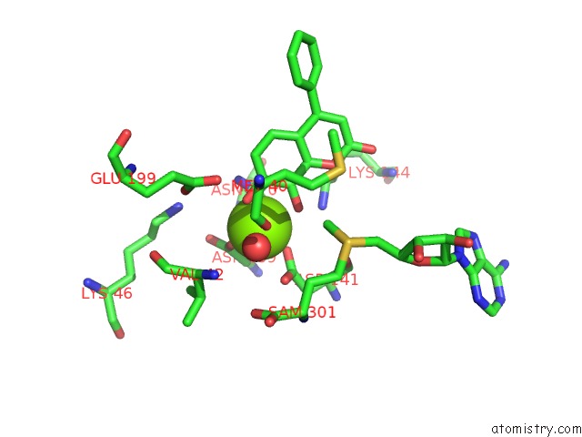
Mono view
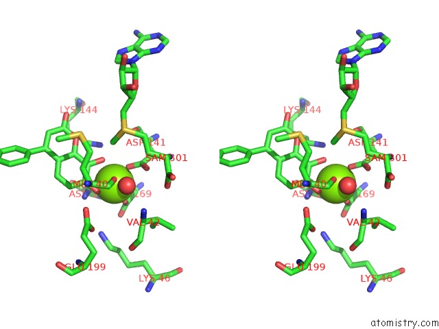
Stereo pair view
Mono view
Stereo pair view
A full contact list of Magnesium with other atoms in the Mg binding
site number 1 of Crystal Structures of Rat Catechol-O-Methyltransferase Complexed with Coumarine-Based Inhibitor within 5.0Å range:
|
Reference:
E.Tsuji,
K.Okazaki,
K.Takeda.
Crystal Structures of Rat Catechol-O-Methyltransferase Complexed with Coumarine-Based Inhibitor Biochem.Biophys.Res.Commun. V. 378 494 2009.
ISSN: ISSN 0006-291X
PubMed: 19056347
DOI: 10.1016/J.BBRC.2008.11.085
Page generated: Sun Aug 10 17:06:03 2025
ISSN: ISSN 0006-291X
PubMed: 19056347
DOI: 10.1016/J.BBRC.2008.11.085
Last articles
Mg in 6FCIMg in 6FCP
Mg in 6FCH
Mg in 6FCD
Mg in 6FCB
Mg in 6FBV
Mg in 6FBP
Mg in 6FBO
Mg in 6FBN
Mg in 6FBE