Magnesium »
PDB 2zws-3a0t »
2zyf »
Magnesium in PDB 2zyf: Crystal Structure of Homocitrate Synthase From Thermus Thermophilus Complexed with Magnesuim Ion and Alpha-Ketoglutarate
Enzymatic activity of Crystal Structure of Homocitrate Synthase From Thermus Thermophilus Complexed with Magnesuim Ion and Alpha-Ketoglutarate
All present enzymatic activity of Crystal Structure of Homocitrate Synthase From Thermus Thermophilus Complexed with Magnesuim Ion and Alpha-Ketoglutarate:
2.3.3.14;
2.3.3.14;
Protein crystallography data
The structure of Crystal Structure of Homocitrate Synthase From Thermus Thermophilus Complexed with Magnesuim Ion and Alpha-Ketoglutarate, PDB code: 2zyf
was solved by
T.Okada,
T.Tomita,
T.Kuzuyama,
M.Nishiyama,
with X-Ray Crystallography technique. A brief refinement statistics is given in the table below:
| Resolution Low / High (Å) | 31.51 / 2.15 |
| Space group | P 63 2 2 |
| Cell size a, b, c (Å), α, β, γ (°) | 135.465, 135.465, 126.612, 90.00, 90.00, 120.00 |
| R / Rfree (%) | 20.3 / 23.2 |
Magnesium Binding Sites:
The binding sites of Magnesium atom in the Crystal Structure of Homocitrate Synthase From Thermus Thermophilus Complexed with Magnesuim Ion and Alpha-Ketoglutarate
(pdb code 2zyf). This binding sites where shown within
5.0 Angstroms radius around Magnesium atom.
In total only one binding site of Magnesium was determined in the Crystal Structure of Homocitrate Synthase From Thermus Thermophilus Complexed with Magnesuim Ion and Alpha-Ketoglutarate, PDB code: 2zyf:
In total only one binding site of Magnesium was determined in the Crystal Structure of Homocitrate Synthase From Thermus Thermophilus Complexed with Magnesuim Ion and Alpha-Ketoglutarate, PDB code: 2zyf:
Magnesium binding site 1 out of 1 in 2zyf
Go back to
Magnesium binding site 1 out
of 1 in the Crystal Structure of Homocitrate Synthase From Thermus Thermophilus Complexed with Magnesuim Ion and Alpha-Ketoglutarate
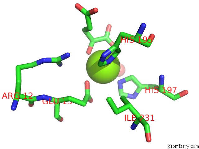
Mono view
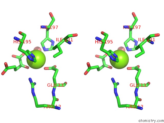
Stereo pair view
Mono view
Stereo pair view
A full contact list of Magnesium with other atoms in the Mg binding
site number 1 of Crystal Structure of Homocitrate Synthase From Thermus Thermophilus Complexed with Magnesuim Ion and Alpha-Ketoglutarate within 5.0Å range:
|
Reference:
T.Okada,
T.Tomita,
A.P.Wulandari,
T.Kuzuyama,
M.Nishiyama.
Mechanism of Substrate Recognition and Insight Into Feedback Inhibition of Homocitrate Synthase From Thermus Thermophilus J.Biol.Chem. 2009.
ISSN: ESSN 1083-351X
PubMed: 19996101
DOI: 10.1074/JBC.M109.086330
Page generated: Wed Aug 14 08:07:19 2024
ISSN: ESSN 1083-351X
PubMed: 19996101
DOI: 10.1074/JBC.M109.086330
Last articles
Zn in 9J0NZn in 9J0O
Zn in 9J0P
Zn in 9FJX
Zn in 9EKB
Zn in 9C0F
Zn in 9CAH
Zn in 9CH0
Zn in 9CH3
Zn in 9CH1