Magnesium »
PDB 2zws-3a0t »
357d »
Magnesium in PDB 357d: 3.5 A Structure of Fragment I From E. Coli 5S Rrna
Protein crystallography data
The structure of 3.5 A Structure of Fragment I From E. Coli 5S Rrna, PDB code: 357d
was solved by
C.C.Correll,
B.Freeborn,
P.B.Moore,
T.A.Steitz,
with X-Ray Crystallography technique. A brief refinement statistics is given in the table below:
| Resolution Low / High (Å) | 10.00 / 3.50 |
| Space group | P 61 2 2 |
| Cell size a, b, c (Å), α, β, γ (°) | 58.670, 58.670, 248.840, 90.00, 90.00, 120.00 |
| R / Rfree (%) | 31.2 / 26.5 |
Other elements in 357d:
The structure of 3.5 A Structure of Fragment I From E. Coli 5S Rrna also contains other interesting chemical elements:
| Mercury | (Hg) | 1 atom |
Magnesium Binding Sites:
The binding sites of Magnesium atom in the 3.5 A Structure of Fragment I From E. Coli 5S Rrna
(pdb code 357d). This binding sites where shown within
5.0 Angstroms radius around Magnesium atom.
In total 8 binding sites of Magnesium where determined in the 3.5 A Structure of Fragment I From E. Coli 5S Rrna, PDB code: 357d:
Jump to Magnesium binding site number: 1; 2; 3; 4; 5; 6; 7; 8;
In total 8 binding sites of Magnesium where determined in the 3.5 A Structure of Fragment I From E. Coli 5S Rrna, PDB code: 357d:
Jump to Magnesium binding site number: 1; 2; 3; 4; 5; 6; 7; 8;
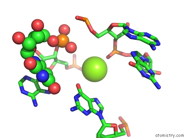
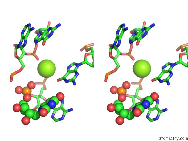
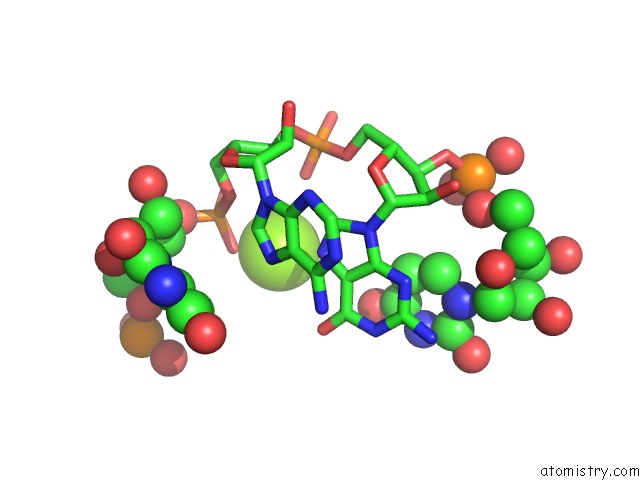
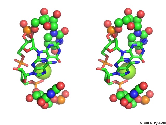
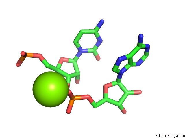
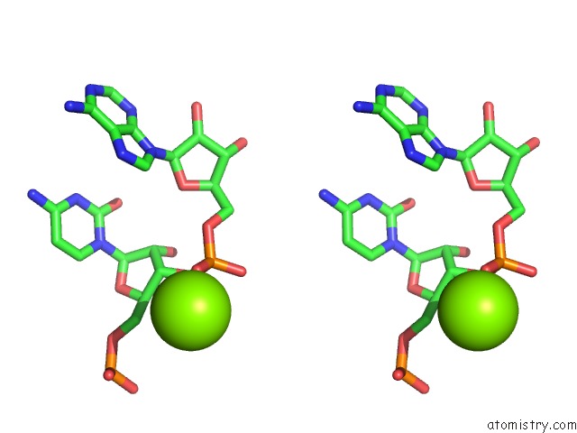
Magnesium binding site 1 out of 8 in 357d
Go back to
Magnesium binding site 1 out
of 8 in the 3.5 A Structure of Fragment I From E. Coli 5S Rrna
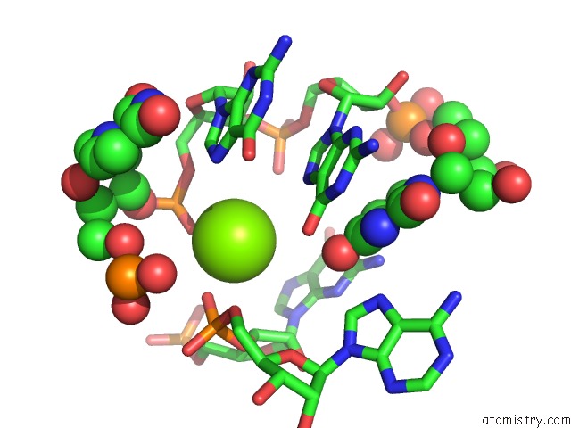
Mono view
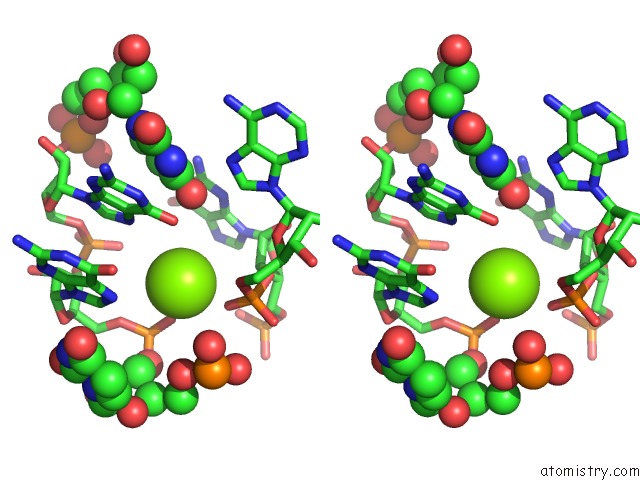
Stereo pair view
Mono view
Stereo pair view
A full contact list of Magnesium with other atoms in the Mg binding
site number 1 of 3.5 A Structure of Fragment I From E. Coli 5S Rrna within 5.0Å range:
|
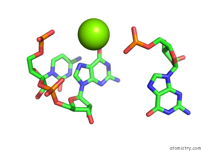
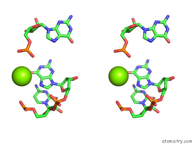
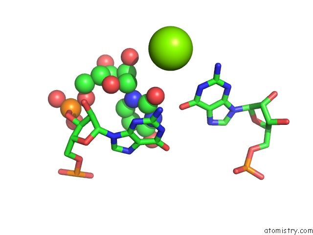
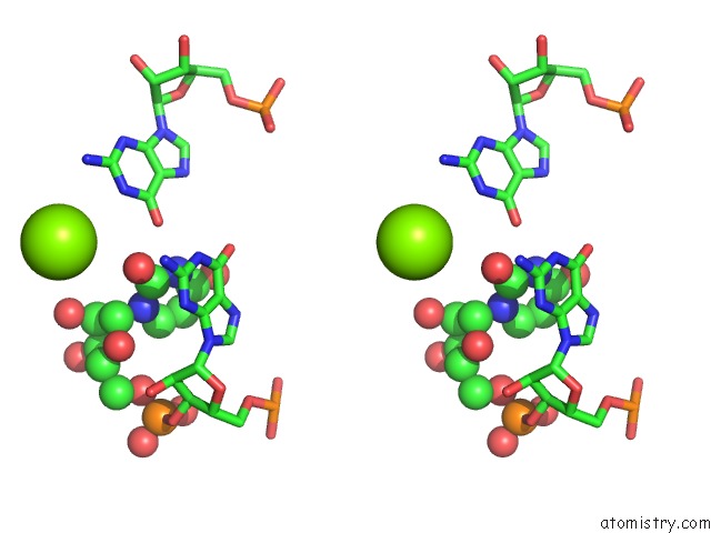
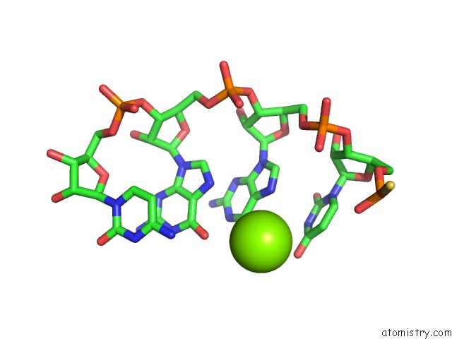
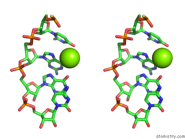
Magnesium binding site 2 out of 8 in 357d
Go back to
Magnesium binding site 2 out
of 8 in the 3.5 A Structure of Fragment I From E. Coli 5S Rrna
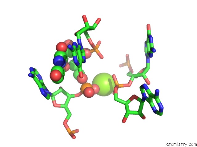
Mono view
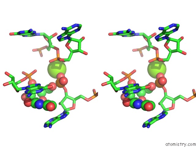
Stereo pair view
Mono view
Stereo pair view
A full contact list of Magnesium with other atoms in the Mg binding
site number 2 of 3.5 A Structure of Fragment I From E. Coli 5S Rrna within 5.0Å range:
|
Magnesium binding site 3 out of 8 in 357d
Go back to
Magnesium binding site 3 out
of 8 in the 3.5 A Structure of Fragment I From E. Coli 5S Rrna
Mono view
Stereo pair view
Mono view
Stereo pair view
A full contact list of Magnesium with other atoms in the Mg binding
site number 3 of 3.5 A Structure of Fragment I From E. Coli 5S Rrna within 5.0Å range:
|
Magnesium binding site 4 out of 8 in 357d
Go back to
Magnesium binding site 4 out
of 8 in the 3.5 A Structure of Fragment I From E. Coli 5S Rrna
Mono view
Stereo pair view
Mono view
Stereo pair view
A full contact list of Magnesium with other atoms in the Mg binding
site number 4 of 3.5 A Structure of Fragment I From E. Coli 5S Rrna within 5.0Å range:
|
Magnesium binding site 5 out of 8 in 357d
Go back to
Magnesium binding site 5 out
of 8 in the 3.5 A Structure of Fragment I From E. Coli 5S Rrna
Mono view
Stereo pair view
Mono view
Stereo pair view
A full contact list of Magnesium with other atoms in the Mg binding
site number 5 of 3.5 A Structure of Fragment I From E. Coli 5S Rrna within 5.0Å range:
|
Magnesium binding site 6 out of 8 in 357d
Go back to
Magnesium binding site 6 out
of 8 in the 3.5 A Structure of Fragment I From E. Coli 5S Rrna
Mono view
Stereo pair view
Mono view
Stereo pair view
A full contact list of Magnesium with other atoms in the Mg binding
site number 6 of 3.5 A Structure of Fragment I From E. Coli 5S Rrna within 5.0Å range:
|
Magnesium binding site 7 out of 8 in 357d
Go back to
Magnesium binding site 7 out
of 8 in the 3.5 A Structure of Fragment I From E. Coli 5S Rrna
Mono view
Stereo pair view
Mono view
Stereo pair view
A full contact list of Magnesium with other atoms in the Mg binding
site number 7 of 3.5 A Structure of Fragment I From E. Coli 5S Rrna within 5.0Å range:
|
Magnesium binding site 8 out of 8 in 357d
Go back to
Magnesium binding site 8 out
of 8 in the 3.5 A Structure of Fragment I From E. Coli 5S Rrna
Mono view
Stereo pair view
Mono view
Stereo pair view
A full contact list of Magnesium with other atoms in the Mg binding
site number 8 of 3.5 A Structure of Fragment I From E. Coli 5S Rrna within 5.0Å range:
|
Reference:
C.C.Correll,
B.Freeborn,
P.B.Moore,
T.A.Steitz.
Metals, Motifs, and Recognition in the Crystal Structure of A 5S Rrna Domain. Cell(Cambridge,Mass.) V. 91 705 1997.
ISSN: ISSN 0092-8674
PubMed: 9393863
DOI: 10.1016/S0092-8674(00)80457-2
Page generated: Wed Aug 14 08:11:07 2024
ISSN: ISSN 0092-8674
PubMed: 9393863
DOI: 10.1016/S0092-8674(00)80457-2
Last articles
Zn in 9MJ5Zn in 9HNW
Zn in 9G0L
Zn in 9FNE
Zn in 9DZN
Zn in 9E0I
Zn in 9D32
Zn in 9DAK
Zn in 8ZXC
Zn in 8ZUF