Magnesium »
PDB 2zws-3a0t »
3a06 »
Magnesium in PDB 3a06: Crystal Structure of Dxr From Thermooga Maritia, in Complex with Fosmidomycin and Nadph
Enzymatic activity of Crystal Structure of Dxr From Thermooga Maritia, in Complex with Fosmidomycin and Nadph
All present enzymatic activity of Crystal Structure of Dxr From Thermooga Maritia, in Complex with Fosmidomycin and Nadph:
1.1.1.267;
1.1.1.267;
Protein crystallography data
The structure of Crystal Structure of Dxr From Thermooga Maritia, in Complex with Fosmidomycin and Nadph, PDB code: 3a06
was solved by
M.Takenoya,
A.Ohtaki,
K.Noguchi,
Y.Sasaki,
K.Ohsawa,
M.Yohda,
S.Yajima,
with X-Ray Crystallography technique. A brief refinement statistics is given in the table below:
| Resolution Low / High (Å) | 40.72 / 2.00 |
| Space group | P 43 |
| Cell size a, b, c (Å), α, β, γ (°) | 108.622, 108.622, 74.662, 90.00, 90.00, 90.00 |
| R / Rfree (%) | 20.7 / 24 |
Magnesium Binding Sites:
The binding sites of Magnesium atom in the Crystal Structure of Dxr From Thermooga Maritia, in Complex with Fosmidomycin and Nadph
(pdb code 3a06). This binding sites where shown within
5.0 Angstroms radius around Magnesium atom.
In total 2 binding sites of Magnesium where determined in the Crystal Structure of Dxr From Thermooga Maritia, in Complex with Fosmidomycin and Nadph, PDB code: 3a06:
Jump to Magnesium binding site number: 1; 2;
In total 2 binding sites of Magnesium where determined in the Crystal Structure of Dxr From Thermooga Maritia, in Complex with Fosmidomycin and Nadph, PDB code: 3a06:
Jump to Magnesium binding site number: 1; 2;
Magnesium binding site 1 out of 2 in 3a06
Go back to
Magnesium binding site 1 out
of 2 in the Crystal Structure of Dxr From Thermooga Maritia, in Complex with Fosmidomycin and Nadph
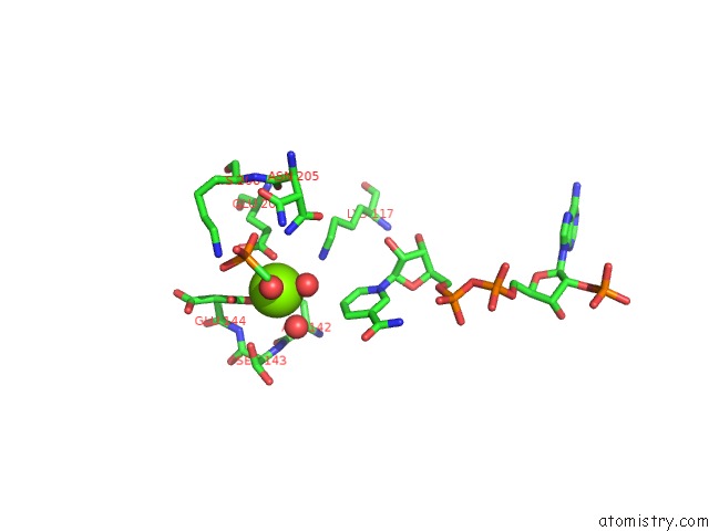
Mono view
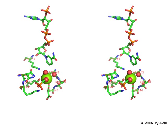
Stereo pair view
Mono view
Stereo pair view
A full contact list of Magnesium with other atoms in the Mg binding
site number 1 of Crystal Structure of Dxr From Thermooga Maritia, in Complex with Fosmidomycin and Nadph within 5.0Å range:
|
Magnesium binding site 2 out of 2 in 3a06
Go back to
Magnesium binding site 2 out
of 2 in the Crystal Structure of Dxr From Thermooga Maritia, in Complex with Fosmidomycin and Nadph
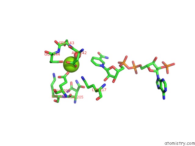
Mono view
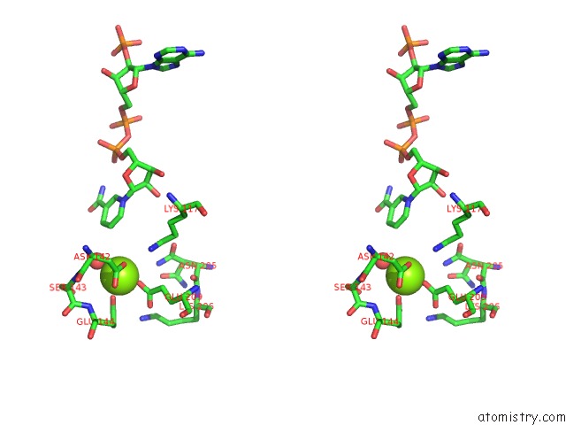
Stereo pair view
Mono view
Stereo pair view
A full contact list of Magnesium with other atoms in the Mg binding
site number 2 of Crystal Structure of Dxr From Thermooga Maritia, in Complex with Fosmidomycin and Nadph within 5.0Å range:
|
Reference:
M.Takenoya,
A.Ohtaki,
K.Noguchi,
K.Endo,
Y.Sasaki,
K.Ohsawa,
S.Yajima,
M.Yohda.
Crystal Structure of 1-Deoxy-D-Xylulose 5-Phosphate Reductoisomerase From the Hyperthermophile Thermotoga Maritima For Insights Into the Coordination of Conformational Changes and An Inhibitor Binding J.Struct.Biol. V. 170 532 2010.
ISSN: ISSN 1047-8477
PubMed: 20353826
DOI: 10.1016/J.JSB.2010.03.015
Page generated: Sun Aug 10 17:11:43 2025
ISSN: ISSN 1047-8477
PubMed: 20353826
DOI: 10.1016/J.JSB.2010.03.015
Last articles
Mg in 4L9ZMg in 4L9Y
Mg in 4LA6
Mg in 4L9W
Mg in 4L81
Mg in 4L9S
Mg in 4L8N
Mg in 4L87
Mg in 4L8G
Mg in 4L80