Magnesium »
PDB 3a0u-3abk »
3a28 »
Magnesium in PDB 3a28: Crystal Structure of L-2,3-Butanediol Dehydrogenase
Protein crystallography data
The structure of Crystal Structure of L-2,3-Butanediol Dehydrogenase, PDB code: 3a28
was solved by
M.Otagiri,
G.Kurisu,
S.Ui,
M.Kusunoki,
with X-Ray Crystallography technique. A brief refinement statistics is given in the table below:
| Resolution Low / High (Å) | 30.00 / 2.00 |
| Space group | P 1 |
| Cell size a, b, c (Å), α, β, γ (°) | 60.800, 69.200, 127.400, 96.10, 100.20, 109.60 |
| R / Rfree (%) | 19.3 / 24 |
Magnesium Binding Sites:
The binding sites of Magnesium atom in the Crystal Structure of L-2,3-Butanediol Dehydrogenase
(pdb code 3a28). This binding sites where shown within
5.0 Angstroms radius around Magnesium atom.
In total 4 binding sites of Magnesium where determined in the Crystal Structure of L-2,3-Butanediol Dehydrogenase, PDB code: 3a28:
Jump to Magnesium binding site number: 1; 2; 3; 4;
In total 4 binding sites of Magnesium where determined in the Crystal Structure of L-2,3-Butanediol Dehydrogenase, PDB code: 3a28:
Jump to Magnesium binding site number: 1; 2; 3; 4;
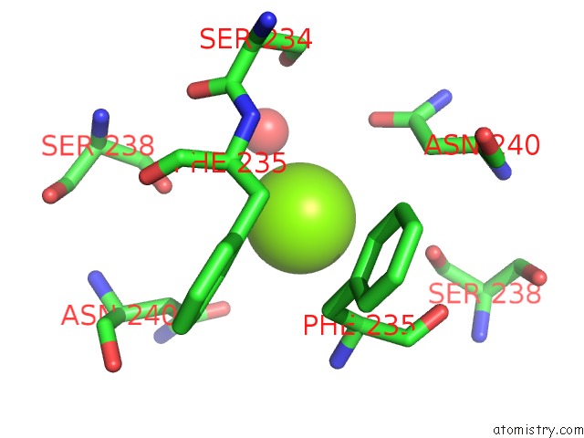
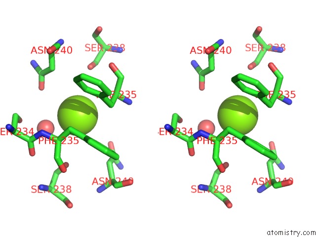
Magnesium binding site 1 out of 4 in 3a28
Go back to
Magnesium binding site 1 out
of 4 in the Crystal Structure of L-2,3-Butanediol Dehydrogenase
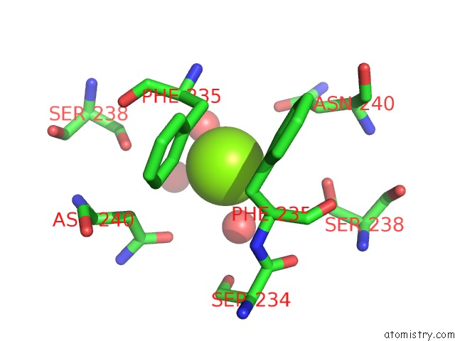
Mono view
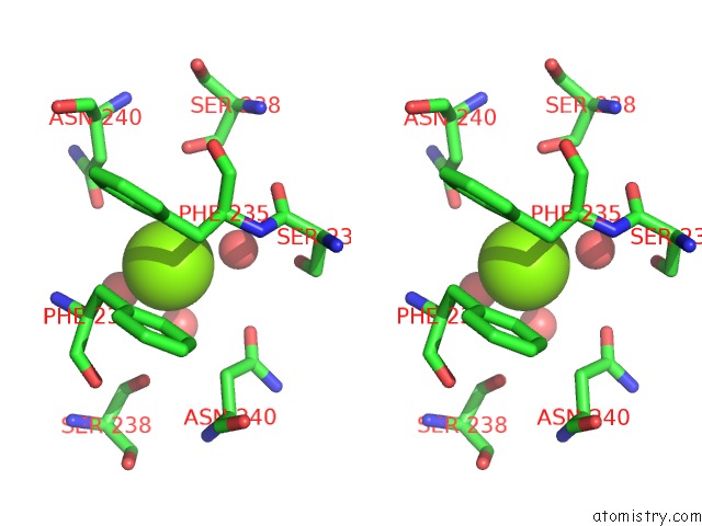
Stereo pair view
Mono view
Stereo pair view
A full contact list of Magnesium with other atoms in the Mg binding
site number 1 of Crystal Structure of L-2,3-Butanediol Dehydrogenase within 5.0Å range:
|
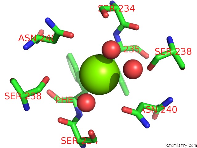
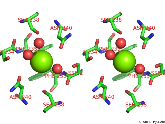
Magnesium binding site 2 out of 4 in 3a28
Go back to
Magnesium binding site 2 out
of 4 in the Crystal Structure of L-2,3-Butanediol Dehydrogenase
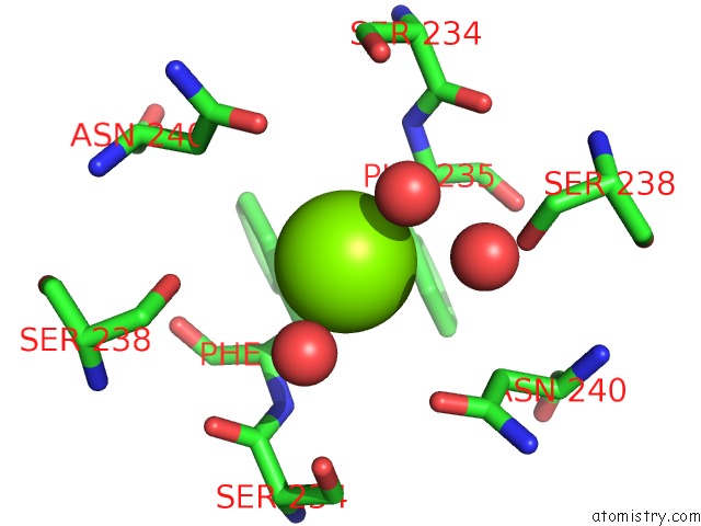
Mono view
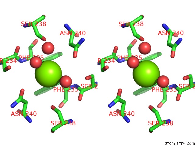
Stereo pair view
Mono view
Stereo pair view
A full contact list of Magnesium with other atoms in the Mg binding
site number 2 of Crystal Structure of L-2,3-Butanediol Dehydrogenase within 5.0Å range:
|
Magnesium binding site 3 out of 4 in 3a28
Go back to
Magnesium binding site 3 out
of 4 in the Crystal Structure of L-2,3-Butanediol Dehydrogenase
Mono view
Stereo pair view
Mono view
Stereo pair view
A full contact list of Magnesium with other atoms in the Mg binding
site number 3 of Crystal Structure of L-2,3-Butanediol Dehydrogenase within 5.0Å range:
|
Magnesium binding site 4 out of 4 in 3a28
Go back to
Magnesium binding site 4 out
of 4 in the Crystal Structure of L-2,3-Butanediol Dehydrogenase
Mono view
Stereo pair view
Mono view
Stereo pair view
A full contact list of Magnesium with other atoms in the Mg binding
site number 4 of Crystal Structure of L-2,3-Butanediol Dehydrogenase within 5.0Å range:
|
Reference:
M.Otagiri,
S.Ui,
Y.Takusagawa,
T.Ohtsuki,
G.Kurisu,
M.Kusunoki.
Structural Basis For Chiral Substrate Recognition By Two 2,3-Butanediol Dehydrogenases Febs Lett. V. 584 219 2010.
ISSN: ISSN 0014-5793
PubMed: 19941855
DOI: 10.1016/J.FEBSLET.2009.11.068
Page generated: Wed Aug 14 08:26:29 2024
ISSN: ISSN 0014-5793
PubMed: 19941855
DOI: 10.1016/J.FEBSLET.2009.11.068
Last articles
Zn in 9J0NZn in 9J0O
Zn in 9J0P
Zn in 9FJX
Zn in 9EKB
Zn in 9C0F
Zn in 9CAH
Zn in 9CH0
Zn in 9CH3
Zn in 9CH1