Magnesium »
PDB 3a0u-3abk »
3a4m »
Magnesium in PDB 3a4m: Crystal Structure of Archaeal O-Phosphoseryl-Trna(Sec) Kinase
Protein crystallography data
The structure of Crystal Structure of Archaeal O-Phosphoseryl-Trna(Sec) Kinase, PDB code: 3a4m
was solved by
Y.Araiso,
R.Ishitani,
D.Soll,
O.Nureki,
with X-Ray Crystallography technique. A brief refinement statistics is given in the table below:
| Resolution Low / High (Å) | 46.50 / 1.79 |
| Space group | P 1 21 1 |
| Cell size a, b, c (Å), α, β, γ (°) | 58.156, 74.524, 64.817, 90.00, 113.37, 90.00 |
| R / Rfree (%) | 20.9 / 25.3 |
Other elements in 3a4m:
The structure of Crystal Structure of Archaeal O-Phosphoseryl-Trna(Sec) Kinase also contains other interesting chemical elements:
| Iodine | (I) | 2 atoms |
Magnesium Binding Sites:
The binding sites of Magnesium atom in the Crystal Structure of Archaeal O-Phosphoseryl-Trna(Sec) Kinase
(pdb code 3a4m). This binding sites where shown within
5.0 Angstroms radius around Magnesium atom.
In total 2 binding sites of Magnesium where determined in the Crystal Structure of Archaeal O-Phosphoseryl-Trna(Sec) Kinase, PDB code: 3a4m:
Jump to Magnesium binding site number: 1; 2;
In total 2 binding sites of Magnesium where determined in the Crystal Structure of Archaeal O-Phosphoseryl-Trna(Sec) Kinase, PDB code: 3a4m:
Jump to Magnesium binding site number: 1; 2;
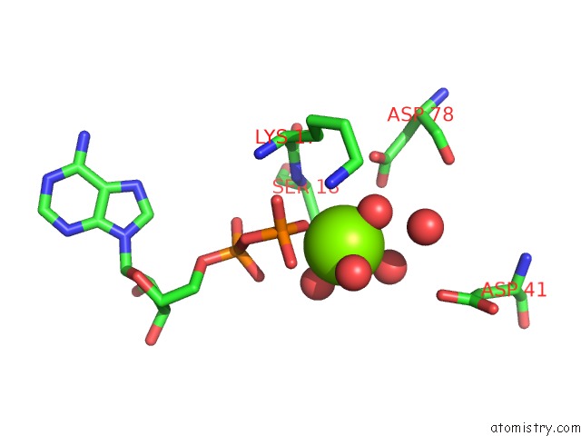
Magnesium binding site 1 out of 2 in 3a4m
Go back to
Magnesium binding site 1 out
of 2 in the Crystal Structure of Archaeal O-Phosphoseryl-Trna(Sec) Kinase

Mono view
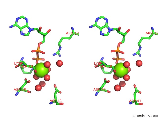
Stereo pair view
Mono view
Stereo pair view
A full contact list of Magnesium with other atoms in the Mg binding
site number 1 of Crystal Structure of Archaeal O-Phosphoseryl-Trna(Sec) Kinase within 5.0Å range:
|
Magnesium binding site 2 out of 2 in 3a4m
Go back to
Magnesium binding site 2 out
of 2 in the Crystal Structure of Archaeal O-Phosphoseryl-Trna(Sec) Kinase
Mono view
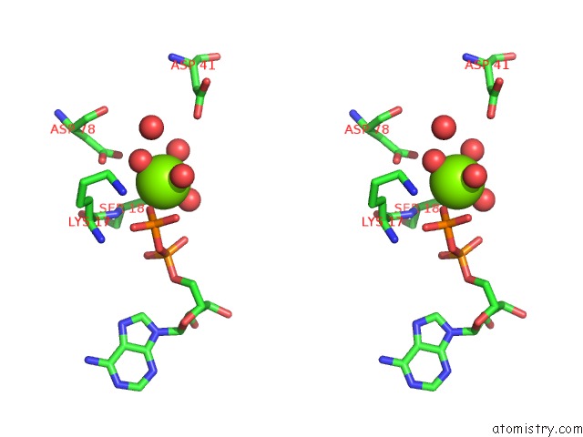
Stereo pair view
Mono view
Stereo pair view
A full contact list of Magnesium with other atoms in the Mg binding
site number 2 of Crystal Structure of Archaeal O-Phosphoseryl-Trna(Sec) Kinase within 5.0Å range:
|
Reference:
Y.Araiso,
R.L.Sherrer,
R.Ishitani,
J.M.L.Ho,
D.Soll,
O.Nureki.
Structure of A Trna-Dependent Kinase Essential For Selenocysteine Decoding Proc.Natl.Acad.Sci.Usa V. 106 16215 2009.
ISSN: ISSN 0027-8424
PubMed: 19805283
DOI: 10.1073/PNAS.0908861106
Page generated: Wed Aug 14 08:27:53 2024
ISSN: ISSN 0027-8424
PubMed: 19805283
DOI: 10.1073/PNAS.0908861106
Last articles
Zn in 9J0NZn in 9J0O
Zn in 9J0P
Zn in 9FJX
Zn in 9EKB
Zn in 9C0F
Zn in 9CAH
Zn in 9CH0
Zn in 9CH3
Zn in 9CH1