Magnesium »
PDB 3a0u-3abk »
3a5k »
Magnesium in PDB 3a5k: Crystal Structure of Protein-Tyrosine Phosphatase 1B
Enzymatic activity of Crystal Structure of Protein-Tyrosine Phosphatase 1B
All present enzymatic activity of Crystal Structure of Protein-Tyrosine Phosphatase 1B:
3.1.3.48;
3.1.3.48;
Protein crystallography data
The structure of Crystal Structure of Protein-Tyrosine Phosphatase 1B, PDB code: 3a5k
was solved by
S.Ito,
with X-Ray Crystallography technique. A brief refinement statistics is given in the table below:
| Resolution Low / High (Å) | 30.51 / 1.85 |
| Space group | P 21 21 21 |
| Cell size a, b, c (Å), α, β, γ (°) | 59.094, 60.755, 87.964, 90.00, 90.00, 90.00 |
| R / Rfree (%) | 18.9 / 23.3 |
Magnesium Binding Sites:
The binding sites of Magnesium atom in the Crystal Structure of Protein-Tyrosine Phosphatase 1B
(pdb code 3a5k). This binding sites where shown within
5.0 Angstroms radius around Magnesium atom.
In total only one binding site of Magnesium was determined in the Crystal Structure of Protein-Tyrosine Phosphatase 1B, PDB code: 3a5k:
In total only one binding site of Magnesium was determined in the Crystal Structure of Protein-Tyrosine Phosphatase 1B, PDB code: 3a5k:
Magnesium binding site 1 out of 1 in 3a5k
Go back to
Magnesium binding site 1 out
of 1 in the Crystal Structure of Protein-Tyrosine Phosphatase 1B
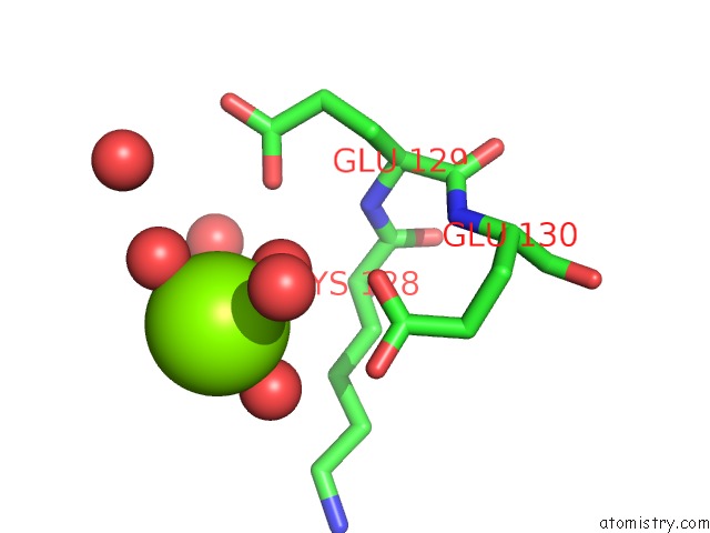
Mono view
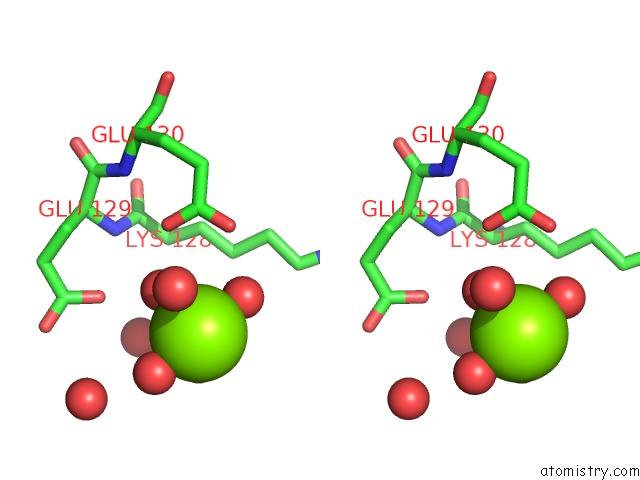
Stereo pair view
Mono view
Stereo pair view
A full contact list of Magnesium with other atoms in the Mg binding
site number 1 of Crystal Structure of Protein-Tyrosine Phosphatase 1B within 5.0Å range:
|
Reference:
N.Iwamoto,
S.Ito,
D.Sumi,
M.Kobayashi,
T.Kumagai.
Post-Translational Modification of A Non-Catalytic CYS121 of PTP1B To Be Published.
Page generated: Sun Aug 10 17:22:50 2025
Last articles
Mg in 3I5XMg in 3I4K
Mg in 3I5F
Mg in 3I5C
Mg in 3I4N
Mg in 3I3E
Mg in 3I4D
Mg in 3I4M
Mg in 3I3D
Mg in 3I3B