Magnesium »
PDB 3a0u-3abk »
3abk »
Magnesium in PDB 3abk: Bovine Heart Cytochrome C Oxidase at the No-Bound Fully Reduced State (50K)
Enzymatic activity of Bovine Heart Cytochrome C Oxidase at the No-Bound Fully Reduced State (50K)
All present enzymatic activity of Bovine Heart Cytochrome C Oxidase at the No-Bound Fully Reduced State (50K):
1.9.3.1;
1.9.3.1;
Protein crystallography data
The structure of Bovine Heart Cytochrome C Oxidase at the No-Bound Fully Reduced State (50K), PDB code: 3abk
was solved by
K.Ohta,
K.Muramoto,
K.Shinzawa-Itoh,
E.Yamashita,
S.Yoshikawa,
T.Tsukihara,
with X-Ray Crystallography technique. A brief refinement statistics is given in the table below:
| Resolution Low / High (Å) | 40.00 / 2.00 |
| Space group | P 21 21 21 |
| Cell size a, b, c (Å), α, β, γ (°) | 182.247, 207.944, 177.997, 90.00, 90.00, 90.00 |
| R / Rfree (%) | 18.3 / 21.9 |
Other elements in 3abk:
The structure of Bovine Heart Cytochrome C Oxidase at the No-Bound Fully Reduced State (50K) also contains other interesting chemical elements:
| Zinc | (Zn) | 2 atoms |
| Iron | (Fe) | 4 atoms |
| Copper | (Cu) | 6 atoms |
| Sodium | (Na) | 2 atoms |
Magnesium Binding Sites:
The binding sites of Magnesium atom in the Bovine Heart Cytochrome C Oxidase at the No-Bound Fully Reduced State (50K)
(pdb code 3abk). This binding sites where shown within
5.0 Angstroms radius around Magnesium atom.
In total 2 binding sites of Magnesium where determined in the Bovine Heart Cytochrome C Oxidase at the No-Bound Fully Reduced State (50K), PDB code: 3abk:
Jump to Magnesium binding site number: 1; 2;
In total 2 binding sites of Magnesium where determined in the Bovine Heart Cytochrome C Oxidase at the No-Bound Fully Reduced State (50K), PDB code: 3abk:
Jump to Magnesium binding site number: 1; 2;
Magnesium binding site 1 out of 2 in 3abk
Go back to
Magnesium binding site 1 out
of 2 in the Bovine Heart Cytochrome C Oxidase at the No-Bound Fully Reduced State (50K)
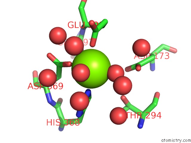
Mono view
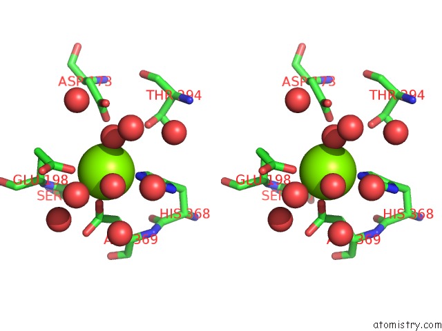
Stereo pair view
Mono view
Stereo pair view
A full contact list of Magnesium with other atoms in the Mg binding
site number 1 of Bovine Heart Cytochrome C Oxidase at the No-Bound Fully Reduced State (50K) within 5.0Å range:
|
Magnesium binding site 2 out of 2 in 3abk
Go back to
Magnesium binding site 2 out
of 2 in the Bovine Heart Cytochrome C Oxidase at the No-Bound Fully Reduced State (50K)
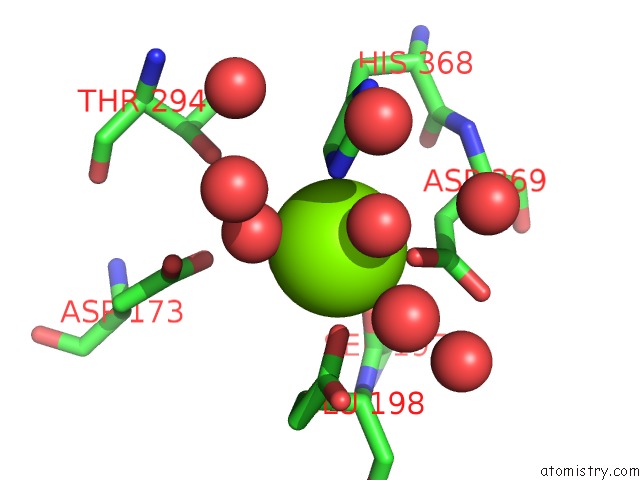
Mono view
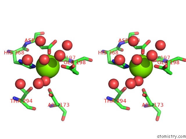
Stereo pair view
Mono view
Stereo pair view
A full contact list of Magnesium with other atoms in the Mg binding
site number 2 of Bovine Heart Cytochrome C Oxidase at the No-Bound Fully Reduced State (50K) within 5.0Å range:
|
Reference:
K.Ohta,
K.Muramoto,
K.Shinzawa-Itoh,
E.Yamashita,
S.Yoshikawa,
T.Tsukihara.
X-Ray Structure of the No-Bound Cu(B) in Bovine Cytochrome C Oxidase Acta Crystallogr.,Sect.F V. 66 251 2010.
ISSN: ESSN 1744-3091
PubMed: 20208153
DOI: 10.1107/S1744309109055109
Page generated: Wed Aug 14 08:31:49 2024
ISSN: ESSN 1744-3091
PubMed: 20208153
DOI: 10.1107/S1744309109055109
Last articles
Zn in 9MJ5Zn in 9HNW
Zn in 9G0L
Zn in 9FNE
Zn in 9DZN
Zn in 9E0I
Zn in 9D32
Zn in 9DAK
Zn in 8ZXC
Zn in 8ZUF