Magnesium »
PDB 3abl-3aln »
3adc »
Magnesium in PDB 3adc: Crystal Structure of O-Phosphoseryl-Trna Kinase Complexed with Selenocysteine Trna and Amppnp (Crystal Type 2)
Protein crystallography data
The structure of Crystal Structure of O-Phosphoseryl-Trna Kinase Complexed with Selenocysteine Trna and Amppnp (Crystal Type 2), PDB code: 3adc
was solved by
Y.Itoh,
S.Chiba,
S.Sekine,
S.Yokoyama,
with X-Ray Crystallography technique. A brief refinement statistics is given in the table below:
| Resolution Low / High (Å) | 28.21 / 2.90 |
| Space group | P 21 21 2 |
| Cell size a, b, c (Å), α, β, γ (°) | 96.824, 263.558, 46.122, 90.00, 90.00, 90.00 |
| R / Rfree (%) | 22.4 / 27.7 |
Magnesium Binding Sites:
The binding sites of Magnesium atom in the Crystal Structure of O-Phosphoseryl-Trna Kinase Complexed with Selenocysteine Trna and Amppnp (Crystal Type 2)
(pdb code 3adc). This binding sites where shown within
5.0 Angstroms radius around Magnesium atom.
In total 2 binding sites of Magnesium where determined in the Crystal Structure of O-Phosphoseryl-Trna Kinase Complexed with Selenocysteine Trna and Amppnp (Crystal Type 2), PDB code: 3adc:
Jump to Magnesium binding site number: 1; 2;
In total 2 binding sites of Magnesium where determined in the Crystal Structure of O-Phosphoseryl-Trna Kinase Complexed with Selenocysteine Trna and Amppnp (Crystal Type 2), PDB code: 3adc:
Jump to Magnesium binding site number: 1; 2;
Magnesium binding site 1 out of 2 in 3adc
Go back to
Magnesium binding site 1 out
of 2 in the Crystal Structure of O-Phosphoseryl-Trna Kinase Complexed with Selenocysteine Trna and Amppnp (Crystal Type 2)
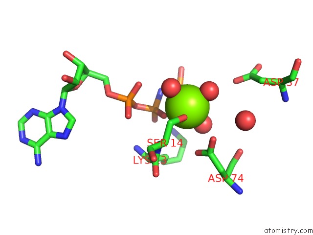
Mono view
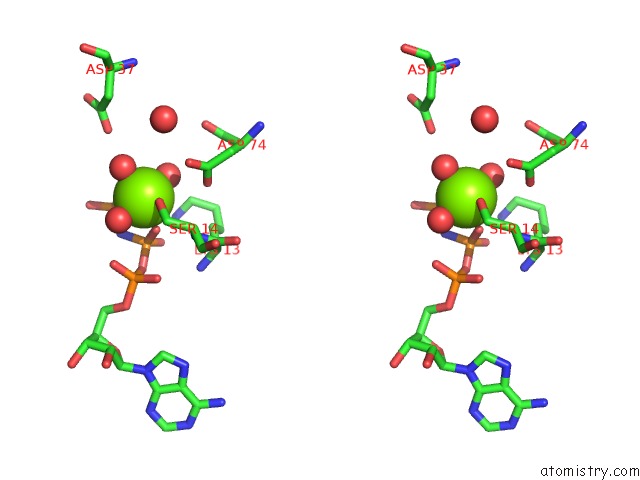
Stereo pair view
Mono view
Stereo pair view
A full contact list of Magnesium with other atoms in the Mg binding
site number 1 of Crystal Structure of O-Phosphoseryl-Trna Kinase Complexed with Selenocysteine Trna and Amppnp (Crystal Type 2) within 5.0Å range:
|
Magnesium binding site 2 out of 2 in 3adc
Go back to
Magnesium binding site 2 out
of 2 in the Crystal Structure of O-Phosphoseryl-Trna Kinase Complexed with Selenocysteine Trna and Amppnp (Crystal Type 2)
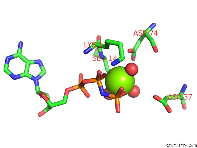
Mono view
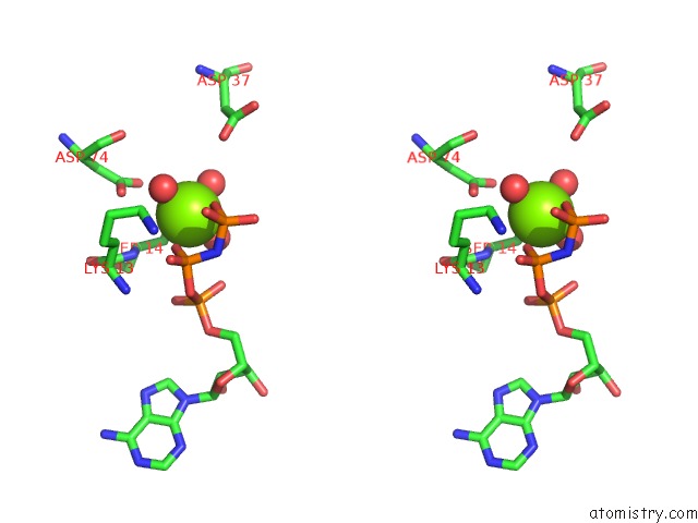
Stereo pair view
Mono view
Stereo pair view
A full contact list of Magnesium with other atoms in the Mg binding
site number 2 of Crystal Structure of O-Phosphoseryl-Trna Kinase Complexed with Selenocysteine Trna and Amppnp (Crystal Type 2) within 5.0Å range:
|
Reference:
S.Chiba,
Y.Itoh,
S.Sekine,
S.Yokoyama.
Structural Basis For the Major Role of O-Phosphoseryl-Trna Kinase in the Uga-Specific Encoding of Selenocysteine Mol.Cell V. 39 410 2010.
ISSN: ISSN 1097-2765
PubMed: 20705242
DOI: 10.1016/J.MOLCEL.2010.07.018
Page generated: Sun Aug 10 17:25:30 2025
ISSN: ISSN 1097-2765
PubMed: 20705242
DOI: 10.1016/J.MOLCEL.2010.07.018
Last articles
Mg in 6C0JMg in 6C06
Mg in 6C05
Mg in 6C04
Mg in 6BZO
Mg in 6BYR
Mg in 6BZ0
Mg in 6BYU
Mg in 6BWH
Mg in 6BWD