Magnesium »
PDB 3abl-3aln »
3agq »
Magnesium in PDB 3agq: Structure of Viral Polymerase Form II
Protein crystallography data
The structure of Structure of Viral Polymerase Form II, PDB code: 3agq
was solved by
D.Takeshita,
K.Tomita,
with X-Ray Crystallography technique. A brief refinement statistics is given in the table below:
| Resolution Low / High (Å) | 20.00 / 3.22 |
| Space group | C 2 2 21 |
| Cell size a, b, c (Å), α, β, γ (°) | 138.769, 255.119, 100.975, 90.00, 90.00, 90.00 |
| R / Rfree (%) | 25.1 / 31.7 |
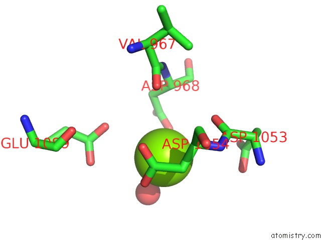
Magnesium Binding Sites:
The binding sites of Magnesium atom in the Structure of Viral Polymerase Form II
(pdb code 3agq). This binding sites where shown within
5.0 Angstroms radius around Magnesium atom.
In total only one binding site of Magnesium was determined in the Structure of Viral Polymerase Form II, PDB code: 3agq:
In total only one binding site of Magnesium was determined in the Structure of Viral Polymerase Form II, PDB code: 3agq:
Magnesium binding site 1 out of 1 in 3agq
Go back to
Magnesium binding site 1 out
of 1 in the Structure of Viral Polymerase Form II
Mono view
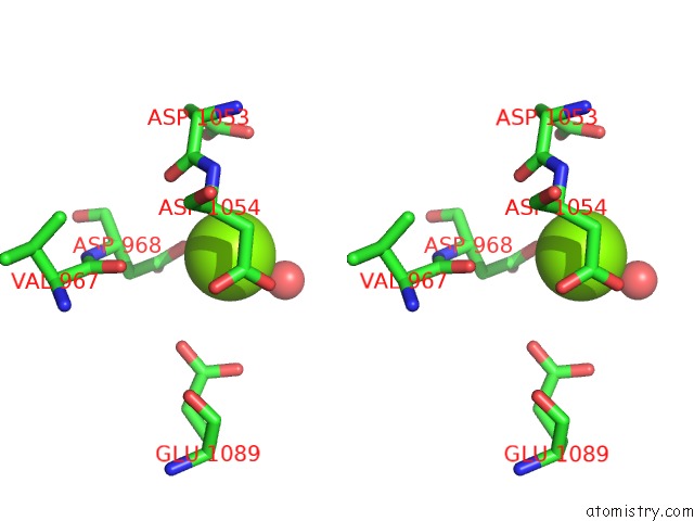
Stereo pair view
Mono view
Stereo pair view
A full contact list of Magnesium with other atoms in the Mg binding
site number 1 of Structure of Viral Polymerase Form II within 5.0Å range:
|
Reference:
D.Takeshita,
K.Tomita.
Assembly of Q{Beta} Viral Rna Polymerase with Host Translational Elongation Factors Ef-Tu and -Ts Proc.Natl.Acad.Sci.Usa V. 107 15733 2010.
ISSN: ISSN 0027-8424
PubMed: 20798060
DOI: 10.1073/PNAS.1006559107
Page generated: Sun Aug 10 17:27:01 2025
ISSN: ISSN 0027-8424
PubMed: 20798060
DOI: 10.1073/PNAS.1006559107
Last articles
Mg in 6K9UMg in 6K8B
Mg in 6K85
Mg in 6K89
Mg in 6K86
Mg in 6K7L
Mg in 6K7M
Mg in 6K7N
Mg in 6K82
Mg in 6K61