Magnesium »
PDB 3abl-3aln »
3ahf »
Magnesium in PDB 3ahf: Phosphoketolase From Bifidobacterium Breve Complexed with Inorganic Phosphate
Enzymatic activity of Phosphoketolase From Bifidobacterium Breve Complexed with Inorganic Phosphate
All present enzymatic activity of Phosphoketolase From Bifidobacterium Breve Complexed with Inorganic Phosphate:
4.1.2.22;
4.1.2.22;
Protein crystallography data
The structure of Phosphoketolase From Bifidobacterium Breve Complexed with Inorganic Phosphate, PDB code: 3ahf
was solved by
R.Suzuki,
T.Katayama,
B.-J.Kim,
T.Wakagi,
H.Shoun,
H.Ashida,
K.Yamamoto,
S.Fushinobu,
with X-Ray Crystallography technique. A brief refinement statistics is given in the table below:
| Resolution Low / High (Å) | 40.88 / 2.30 |
| Space group | I 4 2 2 |
| Cell size a, b, c (Å), α, β, γ (°) | 173.906, 173.906, 163.522, 90.00, 90.00, 90.00 |
| R / Rfree (%) | 17.7 / 22.6 |
Magnesium Binding Sites:
The binding sites of Magnesium atom in the Phosphoketolase From Bifidobacterium Breve Complexed with Inorganic Phosphate
(pdb code 3ahf). This binding sites where shown within
5.0 Angstroms radius around Magnesium atom.
In total only one binding site of Magnesium was determined in the Phosphoketolase From Bifidobacterium Breve Complexed with Inorganic Phosphate, PDB code: 3ahf:
In total only one binding site of Magnesium was determined in the Phosphoketolase From Bifidobacterium Breve Complexed with Inorganic Phosphate, PDB code: 3ahf:
Magnesium binding site 1 out of 1 in 3ahf
Go back to
Magnesium binding site 1 out
of 1 in the Phosphoketolase From Bifidobacterium Breve Complexed with Inorganic Phosphate
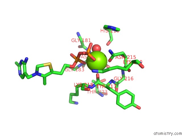
Mono view
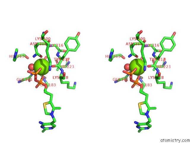
Stereo pair view
Mono view
Stereo pair view
A full contact list of Magnesium with other atoms in the Mg binding
site number 1 of Phosphoketolase From Bifidobacterium Breve Complexed with Inorganic Phosphate within 5.0Å range:
|
Reference:
R.Suzuki,
T.Katayama,
B.-J.Kim,
T.Wakagi,
H.Shoun,
H.Ashida,
K.Yamamoto,
S.Fushinobu.
Crystal Structures of Phosphoketolase: Thiamine Diphosphate-Dependent Dehydration Mechanism J.Biol.Chem. V. 285 34279 2010.
ISSN: ISSN 0021-9258
PubMed: 20739284
DOI: 10.1074/JBC.M110.156281
Page generated: Sun Aug 10 17:27:21 2025
ISSN: ISSN 0021-9258
PubMed: 20739284
DOI: 10.1074/JBC.M110.156281
Last articles
Mg in 6CGDMg in 6CFV
Mg in 6CFU
Mg in 6CFT
Mg in 6CEY
Mg in 6CFS
Mg in 6CFR
Mg in 6CFO
Mg in 6CER
Mg in 6CC3