Magnesium »
PDB 3abl-3aln »
3ai9 »
Magnesium in PDB 3ai9: Crystal Structure of DUF358 Protein Reveals A Putative Spout-Class Rrna Methyltransferase
Protein crystallography data
The structure of Crystal Structure of DUF358 Protein Reveals A Putative Spout-Class Rrna Methyltransferase, PDB code: 3ai9
was solved by
Y.A.Yuan,
H.Y.Chen,
with X-Ray Crystallography technique. A brief refinement statistics is given in the table below:
| Resolution Low / High (Å) | 34.06 / 1.55 |
| Space group | C 1 2 1 |
| Cell size a, b, c (Å), α, β, γ (°) | 84.288, 48.133, 62.362, 90.00, 126.09, 90.00 |
| R / Rfree (%) | 19 / 23.7 |
Magnesium Binding Sites:
The binding sites of Magnesium atom in the Crystal Structure of DUF358 Protein Reveals A Putative Spout-Class Rrna Methyltransferase
(pdb code 3ai9). This binding sites where shown within
5.0 Angstroms radius around Magnesium atom.
In total 2 binding sites of Magnesium where determined in the Crystal Structure of DUF358 Protein Reveals A Putative Spout-Class Rrna Methyltransferase, PDB code: 3ai9:
Jump to Magnesium binding site number: 1; 2;
In total 2 binding sites of Magnesium where determined in the Crystal Structure of DUF358 Protein Reveals A Putative Spout-Class Rrna Methyltransferase, PDB code: 3ai9:
Jump to Magnesium binding site number: 1; 2;
Magnesium binding site 1 out of 2 in 3ai9
Go back to
Magnesium binding site 1 out
of 2 in the Crystal Structure of DUF358 Protein Reveals A Putative Spout-Class Rrna Methyltransferase
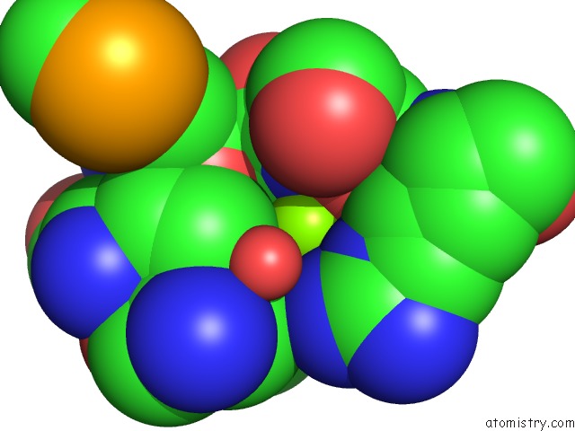
Mono view
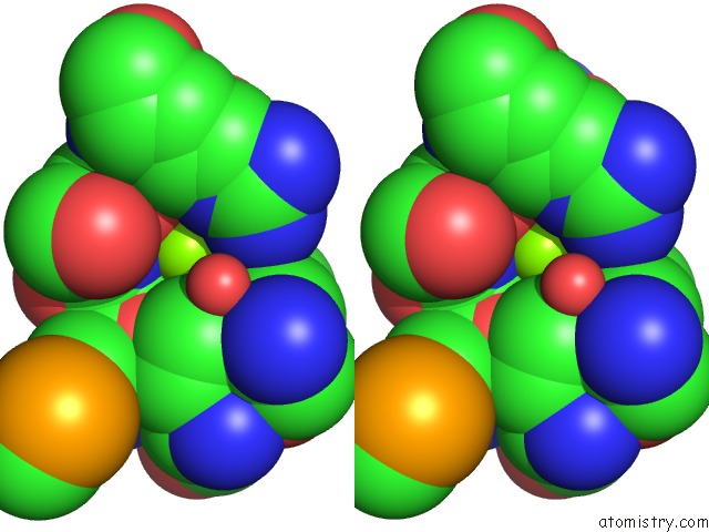
Stereo pair view
Mono view
Stereo pair view
A full contact list of Magnesium with other atoms in the Mg binding
site number 1 of Crystal Structure of DUF358 Protein Reveals A Putative Spout-Class Rrna Methyltransferase within 5.0Å range:
|
Magnesium binding site 2 out of 2 in 3ai9
Go back to
Magnesium binding site 2 out
of 2 in the Crystal Structure of DUF358 Protein Reveals A Putative Spout-Class Rrna Methyltransferase
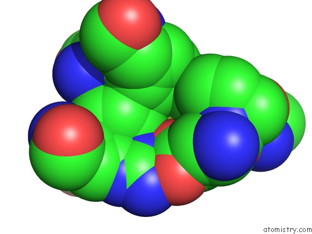
Mono view
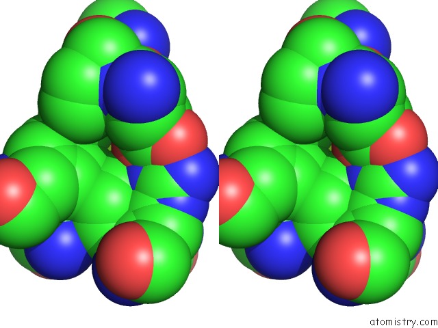
Stereo pair view
Mono view
Stereo pair view
A full contact list of Magnesium with other atoms in the Mg binding
site number 2 of Crystal Structure of DUF358 Protein Reveals A Putative Spout-Class Rrna Methyltransferase within 5.0Å range:
|
Reference:
H.Y.Chen,
Y.A.Yuan.
Crystal Structure of MJ1640/DUF358 Protein Reveals A Putative Spout-Class Rna Methyltransferase J Mol Cell Biol V. 2 366 2010.
ISSN: ISSN 1674-2788
PubMed: 21098051
DOI: 10.1093/JMCB/MJQ034
Page generated: Sun Aug 10 17:27:43 2025
ISSN: ISSN 1674-2788
PubMed: 21098051
DOI: 10.1093/JMCB/MJQ034
Last articles
Mg in 6CAQMg in 6CGD
Mg in 6CFV
Mg in 6CFU
Mg in 6CFT
Mg in 6CEY
Mg in 6CFS
Mg in 6CFR
Mg in 6CFO
Mg in 6CER