Magnesium »
PDB 3abl-3aln »
3al5 »
Magnesium in PDB 3al5: Crystal Structure of Human TYW5
Protein crystallography data
The structure of Crystal Structure of Human TYW5, PDB code: 3al5
was solved by
M.Kato,
Y.Araiso,
R.Ishitani,
O.Nureki,
with X-Ray Crystallography technique. A brief refinement statistics is given in the table below:
| Resolution Low / High (Å) | 50.00 / 2.50 |
| Space group | P 41 21 2 |
| Cell size a, b, c (Å), α, β, γ (°) | 164.942, 164.942, 105.149, 90.00, 90.00, 90.00 |
| R / Rfree (%) | 22 / 28.1 |
Magnesium Binding Sites:
The binding sites of Magnesium atom in the Crystal Structure of Human TYW5
(pdb code 3al5). This binding sites where shown within
5.0 Angstroms radius around Magnesium atom.
In total 2 binding sites of Magnesium where determined in the Crystal Structure of Human TYW5, PDB code: 3al5:
Jump to Magnesium binding site number: 1; 2;
In total 2 binding sites of Magnesium where determined in the Crystal Structure of Human TYW5, PDB code: 3al5:
Jump to Magnesium binding site number: 1; 2;
Magnesium binding site 1 out of 2 in 3al5
Go back to
Magnesium binding site 1 out
of 2 in the Crystal Structure of Human TYW5

Mono view
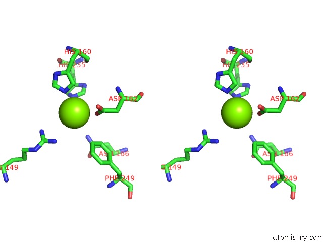
Stereo pair view
Mono view
Stereo pair view
A full contact list of Magnesium with other atoms in the Mg binding
site number 1 of Crystal Structure of Human TYW5 within 5.0Å range:
|
Magnesium binding site 2 out of 2 in 3al5
Go back to
Magnesium binding site 2 out
of 2 in the Crystal Structure of Human TYW5

Mono view
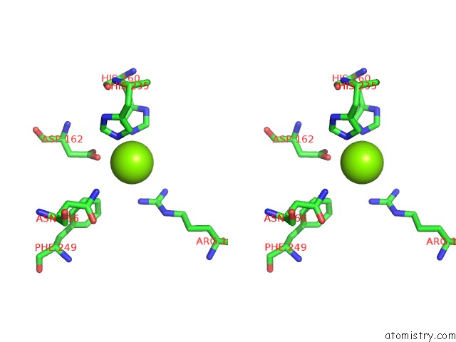
Stereo pair view
Mono view
Stereo pair view
A full contact list of Magnesium with other atoms in the Mg binding
site number 2 of Crystal Structure of Human TYW5 within 5.0Å range:
|
Reference:
M.Kato,
Y.Araiso,
A.Noma,
A.Nagao,
T.Suzuki,
R.Ishitani,
O.Nureki.
Crystal Structure of A Novel Jmjc-Domain-Containing Protein, TYW5, Involved in Trna Modification. Nucleic Acids Res. V. 39 1576 2011.
ISSN: ISSN 0305-1048
PubMed: 20972222
DOI: 10.1093/NAR/GKQ919
Page generated: Sun Aug 10 17:30:52 2025
ISSN: ISSN 0305-1048
PubMed: 20972222
DOI: 10.1093/NAR/GKQ919
Last articles
Mg in 6C62Mg in 6C5U
Mg in 6C5N
Mg in 6C55
Mg in 6C2W
Mg in 6C3P
Mg in 6C3O
Mg in 6C2C
Mg in 6C2X
Mg in 6C25